. 2018 Jun 13;38(24):5478-5494.
doi: 10.1523/JNEUROSCI.3037-17.2018. Epub 2018 May 17.

miR126-5p Downregulation Facilitates Axon Degeneration and NMJ Disruption via a Non-Cell-Autonomous Mechanism in ALS

Affiliations

miR126-5p Downregulation Facilitates Axon Degeneration and NMJ Disruption via a Non-Cell-Autonomous Mechanism in ALS

Roy Maimon et al. J Neurosci. .

Abstract

Axon degeneration and disruption of neuromuscular junctions (NMJs) are key events in amyotrophic lateral sclerosis (ALS) pathology. Although the disease's etiology is not fully understood, it is thought to involve a non-cell-autonomous mechanism and alterations in RNA metabolism. Here, we identified reduced levels of miR126-5p in presymptomatic ALS male mice models, and an increase in its targets: axon destabilizing Type 3 Semaphorins and their coreceptor Neuropilins. Using compartmentalized in vitro cocultures, we demonstrated that myocytes expressing diverse ALS-causing mutations promote axon degeneration and NMJ dysfunction, which were inhibited by applying Neuropilin1 blocking antibody. Finally, overexpressing miR126-5p is sufficient to transiently rescue axon degeneration and NMJ disruption both in vitro and in vivo Thus, we demonstrate a novel mechanism underlying ALS pathology, in which alterations in miR126-5p facilitate a non-cell-autonomous mechanism of motor neuron degeneration in ALS.SIGNIFICANCE STATEMENT Despite some progress, currently no effective treatment is available for amyotrophic lateral sclerosis (ALS). We suggest a novel regulatory role for miR126-5p in ALS and demonstrate, for the first time, a mechanism by which alterations in miR126-5p contribute to axon degeneration and NMJ disruption observed in ALS. We show that miR126-5p is altered in ALS models and that it can modulate Sema3 and NRP protein expression. Furthermore, NRP1 elevations in motor neurons and muscle secretion of Sema3A contribute to axon degeneration and NMJ disruption in ALS. Finally, overexpressing miR126-5p is sufficient to transiently rescue NMJ disruption and axon degeneration both in vitro and in vivo.

Keywords: ALS; NMJ; Sema3A; axon degeneration; miRNA; microfluidic chambers.


Figures

Figure 1.
Figure 1.
Presymptomatic elevation in the levels of Sema3A and NRP1 in ALS models. A, B, Western blot analysis of P30 and P60 GC muscle extracts revealed that the levels of Sema3A are elevated in presymptomatic SOD1G93A muscles compared with their corresponding LM control wherein at earlier stages we found no significant difference (Figure 1-1). Tubulin was used as a loading control. P30 (Student's t test, n = 3, *p = 0.042). P60 (Student's t test, n = 4, *p = 0.038). C, qPCR analysis of presymptomatic P60 and P30 GC muscle extracts also shows an elevation in the mRNA levels of Sema3A in SOD1G93A (Student's t test, SOD1G93A, n = 5, LM, n = 4, *p = 0.049). D, Immunostaining of primary myocytes after 7 d in culture shows elevated levels of Sema3A in primary myocytes of SOD1G93A. White represents Sema3A. Blue represents nuclear DAPI staining. Scale bars, 5 μm. E, Image analysis reveals an increase in Sema3A intensity in SOD1G93A primary myocytes (Student's t test, n = 3, ∼80 cells, ***p = 0.00001). F, Western blot analysis of primary myocyte-CM revealed a higher level of Sema3A in the CM of SOD1G93A. Cultures were lysed after CM collection, and equal loading volumes of lysates were immunoblotted for ERK to validate CM, which was produced from a similar mass of myocytes (Student's t test, n = 3, *p = 0.018). G, H, Immunostaining of fixed whole P60 GC muscles shows distinct Sema3A expression in the NMJs of SOD1G93A mice. White represents Sema3A. Red represents TMR-BTX labeling of acetylcholine receptors on postsynapse. Blue represents presynaptic NFH in neurons. The percentage of muscle fibers expressing Sema3A in their NMJs in P60 SOD1G93A mice is higher (∼100 NMJ per 1 biological repeat; Student's t test, SOD1G93A, n = 4; WT, n = 3; *p = 0.011). Scale bars, 10 μm. We also examined Sema3A expression in later stages of the disease (Figure 1-2). I, Western blot analysis of GC muscle extracts from P60 mice revealed elevated NRP1 levels in the muscles of SOD1G93A. Tubulin was used as a loading control (Student's t test, n = 3, *p = 0.048). J, Western blot analysis of SN extract from P60 mice shows an elevation in the levels of NRP1 in the SNs of SOD1G93A mice (n = 3). K, Western blot analysis of primary MN lysates after 3 d in culture reveals an elevation in the NRP1 levels in SOD1G93A MNs, which are not regulated by Sema3A binding (Figure 1-3). ERK was used as a loading control (Student's t test, n = 3, *p = 0.031). L–N, Immunostaining of primary MNs after 3 d in culture shows an elevation in the levels of NRP1 in both axons (inset, ∼4.1-fold) and somata (∼1.9-fold) of SOD1G93A MNs. White represents NRP1. Blue represents NFH. Somata (Student's t test, n = 3, ∼40 cells, ***p = 0.00021); axons (Student's t test, n = 3, ∼40 cells, *p = 0.012). Scale bars, 10 μm. O, P, Immunostaining of fixed whole P60 GC muscles shows distinct NRP1 expression in the NMJs of SOD1G93A mice. White represents NRP1. Red represents BTX. Blue represents NFH. The percentage of muscle fibers expressing NRP1 in their NMJs in P60 SOD1G93A mice is higher (Student's t test, SOD1G93A, n = 4; WT, n = 3; *p = 0.042). Scale bars, 5 μm. We further examined NRP1 expression in later stages of the disease (Figure 1-2). Elevations in Sema3A and its coreceptor were found also in human sALS samples (Figure 1-4). A–C, E, F, I–K, M, N, Data represent the mean fold difference over the LM control ± SEM.
Figure 2.
Figure 2.
Sema3A as well as primary myocytes expressing diverse ALS-causing mutations impair the growth of wild-type HB9::GFP motor axons and enhance their retraction and degeneration. A, Experimental procedure illustration and representative time-lapse images of HB9::GFP motor axons (Figure 2-1) in the distal compartment of an MFC with no muscles after applying Sema3A to the distal compartment. After 6 h, axons in the distal compartment of chambers that were treated with Sema3A undergo degeneration, whereas axons in the control chamber or axons cotreated with NRP1 antibody and Sema3A continue growing. Scale bar, 20 μm. B, Quantification of the rate of degraded axons in the distal compartment revealed a higher percentage of degradation in chambers that were exposed to Sema3A compared with either control or coapplication of Sema3A and NRP1 antibody (∼60 axons for Sema3A treatment, ∼70 axons for Control; Student's t test; n = 4; mean ± SEM, ***p = 0.00022). C, Schematic view of the experimental procedure in D–F. HB9::GFP SC explants and primary myocytes of SOD1G93A, TDP43A315T, C9orf72-PR50, C9orf72-GR50, or LM, GFP, and SOD1wt as controls were cocultured in an MFC (Figure 2-2), and the growth of HB9::GFP axons was assessed by time-lapse imaging of the microgroove compartment. D, Representative time-lapse images of the HB9::GFP axon growth when cocultured with (left to right) LM, SOD1G93A, and SOD1G93A + NRP1 antibody. The presence of SOD1G93A myocytes in the distal compartment triggers the retraction and degeneration of HB9::GFP motor axons growing in the groove compartment and prevents their traversing. When NRP1 antibody is applied to the distal compartment, together with SOD1G93A-expressing myocytes, axons are less prone to degenerate. Scale bar, 5 μm. E, Quantification of the rate of axons traversing the distal compartment in B shows the mean percentage of axons that traversed the distal compartment out of the total axons in each field (n = 3; NRP1 antibody experiment, n = 4; Student's t test, *p = 0.025, *p = 0.0433). F, Quantification of the rate of axons traversing the distal compartment shows the mean percentage of axons that traversed the distal compartment out of the total number of axons in each field in coculture with TDP43A315T, C9orf72-PR50, C9orf72-GR50 myocytes, or GFP as a control. The traversing rate of HB9::GFP motor axons into the distal compartment in each of the cocultures with muscle-expressing ALS mutations is significantly reduced (n = 3; Student's t test, GR50, *p = 0.0137; PR50, *p = 0.0374; TDP, *p = 0.0304). G, Representative images of fixed and immunostained SOD1G93A motor axons in the distal compartment of an MFC after applying LM or SOD1G93A myocyte CM to the distal compartment. After 48 h, axons in the distal compartment of chambers that were treated with SOD1G93A CM underwent degeneration, whereas axons that were treated with LM CM remained intact. WT MN axons remained intact after application of either CM (Figure 2-3). When NRP1 antibody is applied to the distal compartment, together with SOD1G93A CM, axons are less prone to degenerate, suggesting the involvement of multiple factors (Figure 2-4). Green represents acetylated tubulin. Scale bar, 20 μm. H, Quantification of the rate of degenerated SOD1G93A axons in the distal compartment treated with control CM, SOD1G93A CM, or SOD1G93A CM that was coincubated with anti-NRP1 antibody (Student's t test; n = 3; ***p = 5 × 10⁻⁷, *p = 0.018). **p < 0.01.
Figure 3.
Figure 3.
miR126-5p is depleted in SOD1G93A muscles and regulates Sema3 and NRP expression. A, NanoString chip screen heat map of significantly altered miRs in P60 muscles of SOD1G93A compared with LM mice (extended table under Figure 3-1). Red and green represent a high or low abundance of miRs, respectively. *p < 0.05 (Student's t test; n = 3). B, miR126-5p was the most significantly downregulated miRNA in SOD1G93A muscles (Student's t test; n = 3, **p = 0.003). C, qPCR analysis of P60 GC muscle extracts further validates the decrease in miR 126-5p in SOD1G93A (n = 3). D–G, qPCR analysis of Sema3A, NRP1, Sema3B, and NRP2 transcript levels in HeLa cells overexpressing either miR126-5p or miR142 demonstrates a reduction in their expression levels specifically under miR126-5p overexpression (Student's t test; n = 3, *p = 0.0438, *p = 0.034, *p = 0.031, and *p = 0.0434, respectively). H, Representative TIRF images of U87MG cells reveal a detachment of the cell membrane from the culture dish surface after Sema3A is added to the culture medium. Scale bar, 10 μm. I, Impedance recording of live cells over time shows that U87MG cells overexpressing miR126-5p are unresponsive to Sema3A added to the culture medium because their impedance continuously increases, whereas the impedance of U87MG cells overexpressing miR142 decreases after treatment (Figure 3-2).
Figure 4.
Figure 4.
Overexpression of miR126-5p in primary SOD1G93A myocytes blocks motor axon degeneration and preserves NMJ activity in a compartmental coculture. A, B, Western blot analysis of transfected myocyte extract overexpressing miR126-5p or miR142 and their CM validates miR126-5p as a regulator of Sema3A specifically in muscles. ERK was used as a loading control (Student's t test, n = 3, *p = 0.0499 and *p = 0.05, respectively). C, Schematic view of the experimental procedure in D, E. HB9::GFP SC explants and primary myocytes of SOD1G93A mice were cocultured in a MFC. The growth of the HB9::GFP axons was assessed both by time-lapse imaging of the microgroove compartment and by imaging axons that traversed the distal compartment. D, Representative time-lapse images and quantification of HB9::GFP axon growth when cocultured with SOD1miR126 myocytes (top) or SOD1miR142 myocytes (bottom). SOD1miR126 myocytes in the distal compartment enhanced the axonal traversal of the distal compartment compared with the SOD1miR142 myocytes. The data are shown as the mean rate of axons that traversed the distal compartment out of the total number of axons in each field ± SEM (Student's t test, n = 3, *p = 0.04216). E, Representative time-lapse images and quantification of HB9::GFP axon growth when cocultured with PR50miR126 myocytes (top) or PR50miR142 myocytes (bottom). PR50miR126 myocytes in the distal compartment enhanced the axonal traversal of the distal compartment compared with PR50miR142 myocytes. The data are shown as the mean rate of axons that traversed the distal compartment out of the total number of axons in each field ± SEM (Student's t test, n = 3, ***p = 0.0039). F, Representative myocyte contraction plot showing the bursting contractile behavior of innervated myocytes in vitro. G, Quantification of the percentage of innervated myocytes that contract in a bursting pattern shows a diminished rate of bursting behavior in SOD1G93A myocytes compared with LM controls. SOD1miR126 myocytes show an increase in the rate of bursting myocytes back to the LM levels. The data are shown as the mean percentage of bursting myocytes ± SEM (Student's t test, n = 3, *p = 0.0291, *p = 0.0156, **p = 0.005656). ***p < 0.001.
Figure 5.
Figure 5.
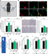
pLL-eGFP-miR126-5p injected into GC muscles of presymptomatic SOD1G93A mice transiently rescues the early phenotype appearance in vivo. A, Schematic view of the in vivo experimental procedure. SOD1G93A mice were injected with either pLL-eGFP-miR126-5p or pLL-eGFP-miR142 in their right or left GC muscles, respectively. Viral infection was validated (Figure 5-1). B, Representative whole-mount NMJ immunostaining of ∼P90 SOD1G93A GC muscles injected with either miR126-5p or miR142 lenti vectors. Red represents BTX. Green represents NFH + synaptophysin in presynaptic neurons. Scale bar, 20 μm. C, The percentage of innervated NMJs in miR126-5p-injected muscles is higher compared with its controls in both P90 and P120 (P90: Student's t test, n = 6, *p = 0.0475, **p = 0.001245; P120: Student's t test, n = 5, *p = 0.043, **p = 0.0096). D, Representative histological images of P120 WT, SOD1G93A, miR126-5p, and miR142 GC muscle cross sections after H&E staining. Scale bar, 100 μm. E, Semiquantification of a GC cross section from D shows a significant increase in the minimal muscle fiber diameter of muscles that were injected with miR126-5p (P120: Student's t test, n = 4, *p = 0.031). F, Illustration of the CatWalk XT gait analysis system that monitors mouse footprints. G, Gait analysis MSI parameter indicates the speed at which the paw loses contact with the surface. The MSI for the P90 miR126-5p-injected limbs was significantly higher than for miR142-injected limbs (Student's t test, *p = 0.0355). H, Gait analysis percentage single-support parameter indicates the relative duration of contact of a single paw on the glass floor. The percentage in which the injected animals were used along the run with a single paw was significantly higher compared with SOD1G93A mice and showed similarity to the WT control (Student's t test, SOD1G93A-injected, ***p = 0.0004; WT-SOD1G93A, ***p = 0.000003). I, Gait analysis base of support parameter indicates the average width between the hindpaws. The base of support of both P90- and P120-injected mice was significant higher compared with SOD1G93A (Student's t test, P90 SOD1G93A-injected, ***p = 0.0000006; WT-SOD1G93A, ***p = 0.000007; P120 SOD1G93A-injected, ***p = 0. 0.00003; WT-SOD1G93A, ***p = 0.000000009).
Figure 6.
Figure 6.
Alterations in Semaphorin3A regulation by miR126-5p trigger MN degeneration in ALS. miR126-5p is a negative regulator of Sema3 signaling in skeletal muscles. Downregulation of miR126-5p in ALS disease drives the overexpression and secretion of Sema3A and potentially other NMJ-destabilizing factors in skeletal muscles. The downregulation in miR126-5p in diseased MNs drives the overexpression of NRP1 specifically in axons. The excess binding and activation of the NRP1 receptor by its overexpressed ligand Sema3A as a result of miR126-5p alteration promote NMJ disruption and axon degeneration in a spatially confined process.

References

    1. Arbour D, Tremblay E, Martineau É, Julien JP, Robitaille R (2015) Early and persistent abnormal decoding by glial cells at the neuromuscular junction in an ALS model. J Neurosci 35:688–706. 10.1523/JNEUROSCI.1379-14.2015 - DOI - PMC - PubMed
    1. Ben-Zvi A, Yagil Z, Hagalili Y, Klein H, Lerman O, Behar O (2006) Semaphorin 3A and neurotrophins: a balance between apoptosis and survival signaling in embryonic DRG neurons. J Neurochem 96:585–597. 10.1111/j.1471-4159.2005.03580.x - DOI - PubMed
    1. Ben-Zvi A, Manor O, Schachner M, Yaron A, Tessier-Lavigne M, Behar O (2008) The semaphorin receptor PlexinA3 mediates neuronal apoptosis during dorsal root ganglia development. J Neurosci 28:12427–12432. 10.1523/JNEUROSCI.3573-08.2008 - DOI - PMC - PubMed
    1. Birger A, Besser E, Reubinoff B, Behar O (2015) A new impedance based approach to test the activity of recombinant protein-semaphorins as a test case. Eur J Cell Biol 94:453–457. 10.1016/j.ejcb.2015.06.006 - DOI - PubMed
    1. Birger A, Ottolenghi M, Perez L, Reubinoff B, Behar O (2018) ALS-related human cortical and motor neurons survival is differentially affected by Sema3A. Cell Death Dis 9:256. 10.1038/s41419-018-0294-6 - DOI - PMC - PubMed

Publication types

MeSH terms

LinkOut - more resources