. 2018 May 17;8(1):7794.
doi: 10.1038/s41598-018-26093-1.

DNAJC17 is localized in nuclear speckles and interacts with splicing machinery components

Affiliations

DNAJC17 is localized in nuclear speckles and interacts with splicing machinery components

A Pascarella et al. Sci Rep. .

Abstract

DNAJC17 is a heat shock protein (HSP40) family member, identified in mouse as susceptibility gene for congenital hypothyroidism. DNAJC17 knockout mouse embryos die prior to implantation. In humans, germline homozygous mutations in DNAJC17 have been found in syndromic retinal dystrophy patients, while heterozygous mutations represent candidate pathogenic events for myeloproliferative disorders. Despite widespread expression and involvement in human diseases, DNAJC17 function is still poorly understood. Herein, we have investigated its function through high-throughput transcriptomic and proteomic approaches. DNAJC17-depleted cells transcriptome highlighted genes involved in general functional categories, mainly related to gene expression. Conversely, DNAJC17 interactome can be classified in very specific functional networks, with the most enriched one including proteins involved in splicing. Furthermore, several splicing-related interactors, were independently validated by co-immunoprecipitation and in vivo co-localization. Accordingly, co-localization of DNAJC17 with SC35, a marker of nuclear speckles, further supported its interaction with spliceosomal components. Lastly, DNAJC17 up-regulation enhanced splicing efficiency of minigene reporter in live cells, while its knockdown induced perturbations of splicing efficiency at whole genome level, as demonstrated by specific analysis of RNAseq data. In conclusion, our study strongly suggests a role of DNAJC17 in splicing-related processes and provides support to its recognized essential function in early development.


Conflict of interest statement

The authors declare no competing interests.

Figures

Figure 1
Figure 1
Gene expression profile analysis of DNAJC17-depleted cells. (A) HeLa cells were transfected with either siRNA specific for DNAJC17 (DNAJC17kd) or scramble (DNAJC17wt). Western blot analysis at 96 h after transfection revealed the specific decline of DNAJC17 (arrow) only in DNAJC17kd cells. Asterisk indicates the aspecific band recognized by the antibody. Tubulin was used as normalization. (B) DNAJC17kd transcriptome was compared with that of DNAJC17wt cells. Pie chart shows that, among the 884 regulated genes, 524 were down-regulated and 360 were up-regulated. (C) Gene Ontology functional analysis showing Biological Process (upper) and Molecular Function (lower) terms significantly enriched in deregulated genes. Enriched terms are represented in dotplot by Count (the number of deregulated genes annotated with the term), p-adjust (corrected p-value of hypergeometric test) and GeneRatio (the ratio between Count and the number of all genes annotated with the term).
Figure 2
Figure 2
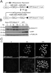
Schematic representation of the Tet-inducible GFP-DNAJC17 fusion protein. (A) The binding of tetracycline to the Tet repressor activates the promoter controlling GFP-DNAJC17 expression. (B) Western blot analysis of whole HeLa (lane 1), GFP-DNAJC17 (lanes 2–4) and GFP (lanes 5–7) cells extracts, showing the expression of GFP-DNAJC17 in a tetracycline dose-dependent manner. GFP-DNAJC17 (upper panel) and endogenous DNAJC17 (second panel) have been revealed by the same western blot with DNAJC17 antibody, captured at lower or higher exposure time, respectively. Molecular mass of each detected protein is indicated on the left. (C) Immunofluorescence at the same time points as (B) shows that GFP-DNAJC17 localizes in the nucleus, while GFP alone localize also in the cytoplasm.
Figure 3
Figure 3
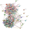
Map of the Interactome Network showing 70 proteins interacting with DNAJC17 protein. In particular, the reported interactors of the GFP-DNAJC17 fusion protein are those remaining after subtraction of GFP counterpart interactors. According to their cellular function, most of the proteins shown in this network were classified into two general groups: spliceosome (top right image) and ribosome (lower left image).
Figure 4
Figure 4
Validation of DNAJC17-interacting partners. Lysates from GFP-DNAJC17 and GFP cells were subjected to co-immunoprecipitation analysis with GFP-Trap®_A affinity beads (Chromotek). Immunoprecipitates were analyzed by Western blotting with the antibodies indicated on the right of each image. The image shows that DNAJC17 co-immunoprecipitates with SNRNP200, PRP19, PLRG-1, CDC5L and XAB2. Molecular mass of each detected protein is indicated on the left.
Figure 5
Figure 5
Confocal analysis of GFP-DNAJC17 cells stained with PRP19, CDC5L and SNRNP200 antibodies. GFP-DNAJC17 cells were induced with tetracycline for 16 h. Endogenous GFP-DNAJC17 fluorescence is shown in green. Staining with anti-PRP19, anti-CDC5L, anti-SNRNP200 or anti-PLRG1 is shown in red. The last column shows a detail of the merged image (right panel). Co-localization is represented with increasing intensity of yellow in the merged images and their details (scale bar 10 µm).
Figure 6
Figure 6
Confocal analysis of GFP-DNAJC17 cells stained with PRP19 and SC35 antibodies. GFP-DNAJC17 cells were induced with tetracycline for 16 h. Endogenous GFP-DNAJC17 fluorescence is shown in green. (A) Staining with anti-SC35 is shown in red. The last panel shows a detail of the merged image (right image). Co-localization is represented with increasing intensity of yellow in the third and last images (B). Anti-SC35 staining is shown in red, while the anti-PRP19 is shown in blue. Co-localization is represented with increasing intensity of pink in the fourth and last images (scale bar 10 µm).
Figure 7
Figure 7
DNAJC17 regulates RNA splicing. (A) Schematic representation of E1A-derived splicing variants. (B) Splicing of E1A was analyzed by RT–PCR in E1A minigene-transfected cells. GFP cells (lanes 1 and 2) and GFP- DNAJC17 cells (lanes 3 and 4) were untreated or treated with Tet for 6 h. Splicing products are indicated on the right of the panel. (C) Quantification of the major E1A mRNA variants: white bars and black bars represent the percentage of each isoform in GFP and GFP-DNAJC17 cells, respectively, either untreated or treated with Tet for 6 h. Data represent the mean ± SD of three independent experiments.
Figure 8
Figure 8
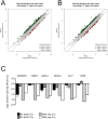
Splicing efficiency in DNAJC17kd. The scatter plots represents for each intron 5′ (A) and 3′ (B) splicing efficiency in DNAJC17kd (x axis) and in the control DNAJC17wt (y axis), as the mean across the three replicates. Considering a cutoff of absolute values ≥1, introns whose splicing efficiency is reduced following DNAJC17 silencing are labeled in green; instead red indicates increased efficiency compared with control. (C) Comparison of relative splicing efficiencies at the 5′ and 3′ ends of selected introns calculated from the RNA-seq data with corresponding splicing efficiencies as determined by RT-qPCR (means of 3 independent RT-qPCR experiments ± SD in triplicates). For each intron, ratio between exon-exon junction transreads and intron 5′ or 3′ ends in DNAJC17kd cells are reported relative to DNAJC17wt control.

References

    1. Kampinga HH, Craig EA. The HSP70 chaperone machinery: J proteins as drivers of functional specificity. Nat Rev Mol Cell Biol. 2010;11:579–592. doi: 10.1038/nrm2941. - DOI - PMC - PubMed
    1. Amendola E, et al. A mouse model demonstrates a multigenic origin of congenital hypothyroidism. Endocrinology. 2005;146:5038–5047. doi: 10.1210/en.2005-0882. - DOI - PubMed
    1. Amendola E, et al. A locus on mouse chromosome 2 is involved in susceptibility to congenital hypothyroidism and contains an essential gene expressed in thyroid. Endocrinology. 2010;151:1948–1958. doi: 10.1210/en.2009-1240. - DOI - PubMed
    1. Patel N, et al. Expanding the clinical, allelic, and locus heterogeneity of retinal dystrophies. Genet Med. 2016;18:554–562. doi: 10.1038/gim.2015.127. - DOI - PubMed
    1. Al Assaf C, et al. Screening of JAK2 V617F and MPL W515 K/L negative essential thrombocythaemia patients for mutations in SESN2, DNAJC17, ST13, TOP1MT, and NTRK1. Br J Haematol. 2014;165:734–737. doi: 10.1111/bjh.12790. - DOI - PMC - PubMed

Publication types

Substances

LinkOut - more resources