. 2018 May 3:9:429.
doi: 10.3389/fphar.2018.00429. eCollection 2018.

The Cytotoxic Effects of Betulin-Conjugated Gold Nanoparticles as Stable Formulations in Normal and Melanoma Cells

Affiliations

The Cytotoxic Effects of Betulin-Conjugated Gold Nanoparticles as Stable Formulations in Normal and Melanoma Cells

Marius Mioc et al. Front Pharmacol. .

Abstract

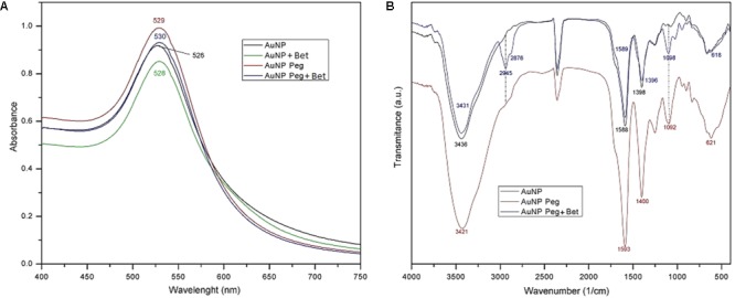
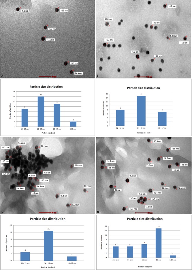
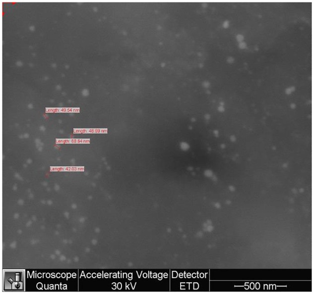
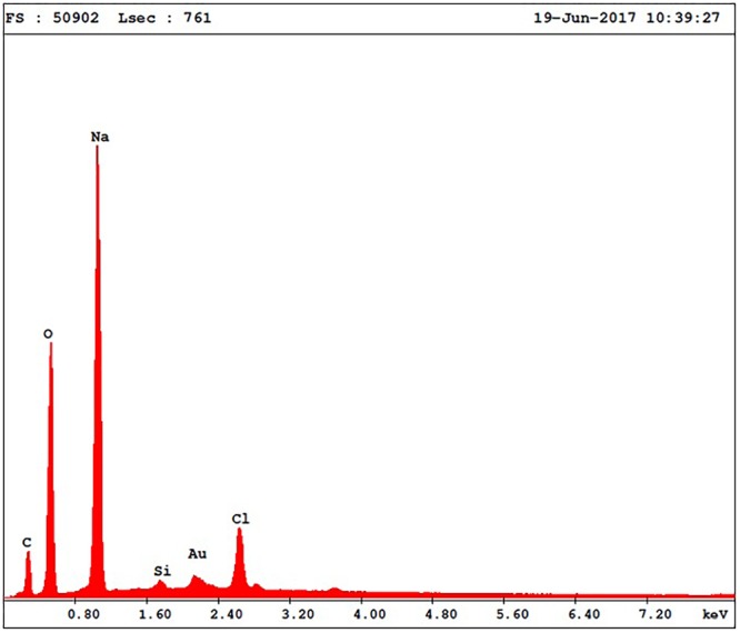
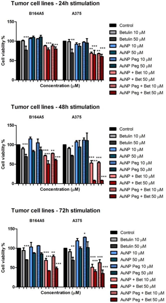
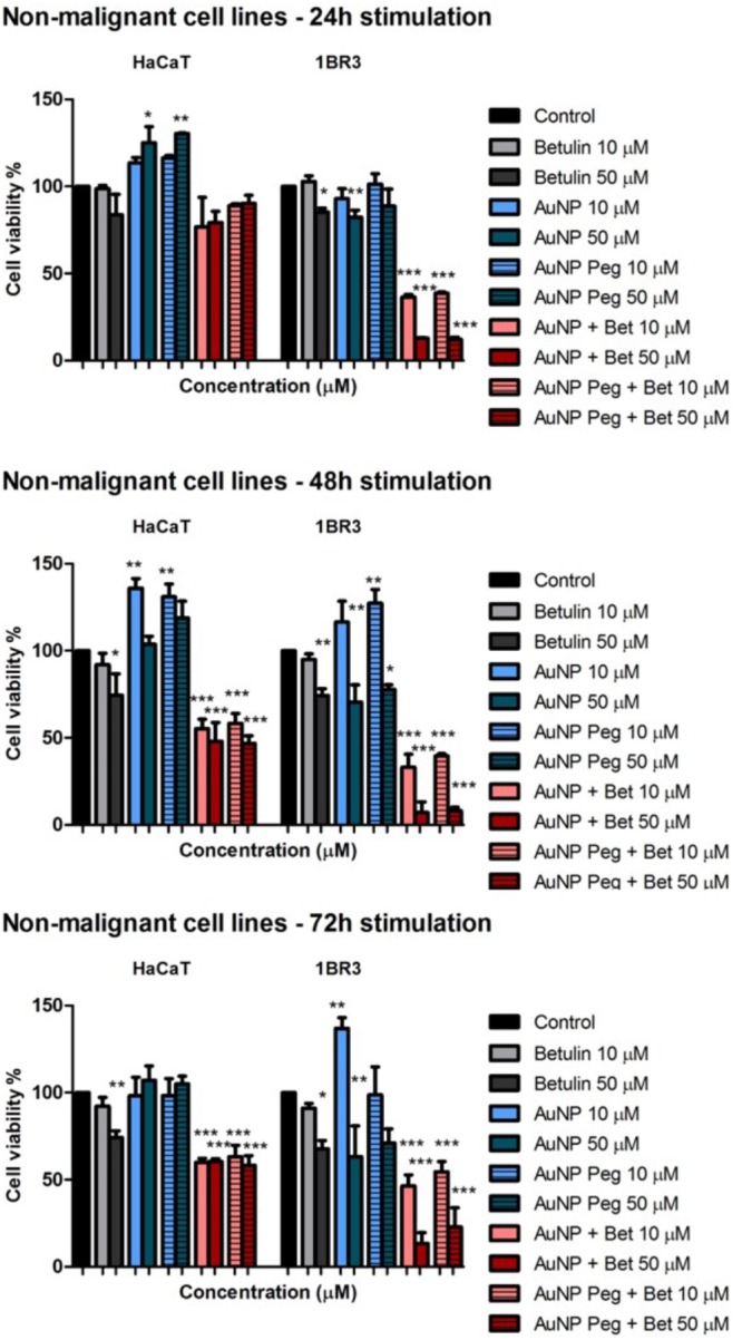
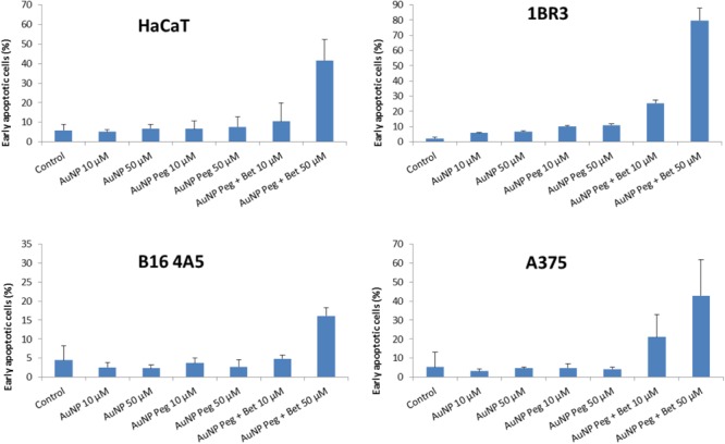
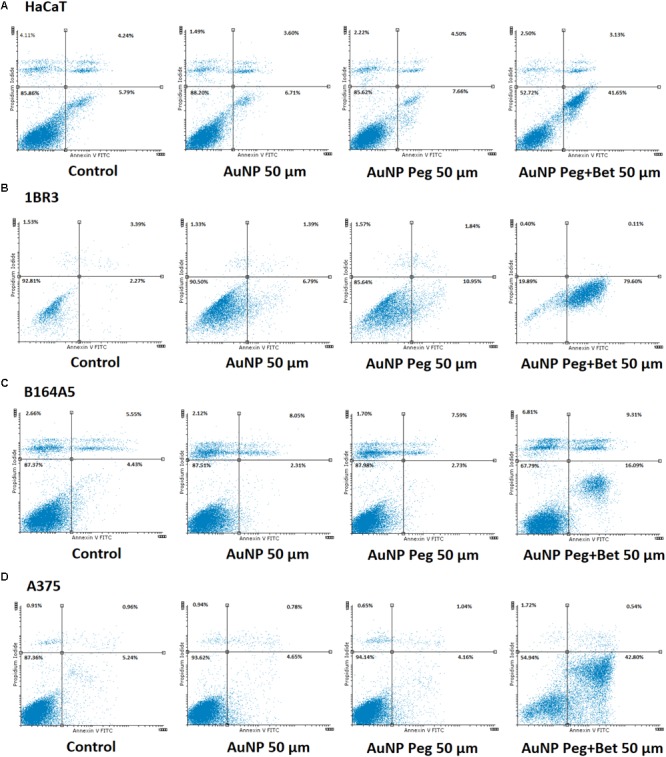
Gold nanoparticles are currently investigated as theranostics tools in cancer therapy due to their proper biocompatibility and increased efficacy related to the ease to customize the surface properties and to conjugate other molecules. Betulin, [lup-20(29)-ene-3β, 28-diol], is a pentacyclic triterpene that has raised scientific interest due to its antiproliferative effect on several cancer types. Herein we described the synthesis of surface modified betulin-conjugated gold nanoparticles using a slightly modified Turkevich method. Transmission electron microscopy (TEM) imaging, dynamic light scattering (DLS), scanning electron microscopy (SEM) and energy-dispersive X-ray spectroscopy (EDX) were used for the characterization of obtained gold nanoparticles. Cytotoxic activity and apoptosis assessment were carried out using the MTT and Annexin V/PI apoptosis assays. The in vitro results showed that betulin coated gold nanoparticles presented a dose-dependent cytotoxic effect and induced apoptosis in all tested cell lines.

Keywords: apoptosis; betulin; gold nanoparticles; melanoma; skin.


Figures

FIGURE 1
FIGURE 1
Recorded UV-Vis spectra for AuNP formulations: AuNP, AuNP + Bet, AuNP Peg and AuNP Peg + Bet (A) and FTIR spectra of AuNP formulations (B).
FIGURE 2
FIGURE 2
Transmission electron microscopy (TEM) analysis and particle size distribution for nanogold samples: AuNP (A), AuNP Peg (B), AuNP + Bet (C), AuNP Peg + Bet (D).
FIGURE 3
FIGURE 3
Scanning electron microscopy (SEM) analysis of gold nanoparticles sample (AuNP).
FIGURE 4
FIGURE 4
EDX spectra of gold nanoparticles sample (AuNP).
FIGURE 5
FIGURE 5
In vitro cytotoxicity assessment of naked AuNPs/AuNP Peg and betulin conjugated gold nanoparticles (10 and 50 μM) on tumor cells: murine – B164A5 and human – A375 melanoma cells at 24, 48, and 72 h post-stimulation by the means of MTT assay. The results are expressed as cell viability percentage (%) related to control cells (stimulated with dimethyl sulfoxide – DMSO). The data represent the mean values ± SD of three independent experiments performed in triplicate. One-way ANOVA analysis was applied to determine the statistical differences followed by Tukey post-test (p < 0.05; ∗∗p < 0.01; ∗∗∗p < 0.001).
FIGURE 6
FIGURE 6
In vitro cytotoxicity assessment of naked AuNPs/AuNP Peg and betulin conjugated gold nanoparticles (10 and 50 μM) on non-malignant human cells: keratinocytes – HaCat and fibroblasts – 1BR3 melanoma cells at 24, 48, and 72 h post-stimulation by the means of MTT assay. The results are expressed as cell viability percentage (%) related to control cells (stimulated with dimethyl sulfoxide – DMSO). The data represent the mean values ± SD of three independent experiments performed in triplicate. One-way ANOVA analysis was applied to determine the statistical differences followed by Tukey post-test (p < 0.05; ∗∗p < 0.01; ∗∗∗p < 0.001).
FIGURE 7
FIGURE 7
Summarized results of early apoptotic cells percentage of HaCaT, 1BR3, B16 4A5 and A375 cell lines after 72 h of treatment with the tested substances. The results are representative of three independent experiments in triplicate and expressed as mean ± SD.
FIGURE 8
FIGURE 8
Representative dotplots of apoptotic cells stained with Annexin V-FITC/PI and analyzed by flow cytometry after 72 h treatment, grouped as follows: (A) HaCat: Control, AuNP 50 μM, AuNP Peg 50 μM, AuNP Peg + Bet 50 μM; (B) 1BR3: Control, AuNP 50 μM, AuNP Peg 50 μM, AuNP Peg + Bet 50 μM; (C) B164A5: Control, AuNP 50 μM, AuNP Peg 50 μM, AuNP Peg + Bet 50 μM; and (D) A375: Control, AuNP 50 μM, AuNP Peg 50 μM, AuNP Peg + Bet 50 μM. Selection of the dotplots was based on the significant pro-apoptotic effects induced by test compounds at 50 μM.

References

    1. Agunloye E., Gavriilidis A., Mazzei L. (2017). A mathematical investigation of the Turkevich organizer theory in the citrate method for the synthesis of gold nanoparticles. Chem. Eng. Sci. 173 275–286. 10.1016/j.ces.2017.07.032 - DOI
    1. Ahmad M. Z., Akhter S., Jain G. K., Rahman M., Pathan S. A., Ahmad F. J., et al. (2010). Metallic nanoparticles: technology overview & drug delivery applications in oncology. Expert Opin. Drug Deliv. 7 927–942. 10.1517/17425247.2010.498473 - DOI - PubMed
    1. Alakurtti S., Makela T., Koskimies S., Yli-Kauhaluoma J. (2006). Pharmacological properties of the ubiquitous natural product betulin. Eur. J. Pharm. Sci. 29 1–13. 10.1016/j.ejps.2006.04.006 - DOI - PubMed
    1. Alkilany A. M., Bani Yaseen A. I., Kailani M. H. (2015). Synthesis of monodispersed gold nanoparticles with exceptional colloidal stability with grafted polyethylene glycol-g-polyvinyl alcohol. J. Nanomater. 16:51 10.1155/2015/712359 - DOI
    1. Arvizo R., Bhattacharya R., Mukherjee P. (2010). Gold nanoparticles: opportunities and challenges in nanomedicine. Expert Opin. Drug Deliv. 7 753–763. 10.1517/17425241003777010 - DOI - PMC - PubMed