. 2018 Apr 15;8(10):2814-2829.
doi: 10.7150/thno.23012. eCollection 2018.

Long noncoding RNA miR503HG, a prognostic indicator, inhibits tumor metastasis by regulating the HNRNPA2B1/NF-κB pathway in hepatocellular carcinoma

Affiliations

Long noncoding RNA miR503HG, a prognostic indicator, inhibits tumor metastasis by regulating the HNRNPA2B1/NF-κB pathway in hepatocellular carcinoma

Hui Wang et al. Theranostics. .

Abstract

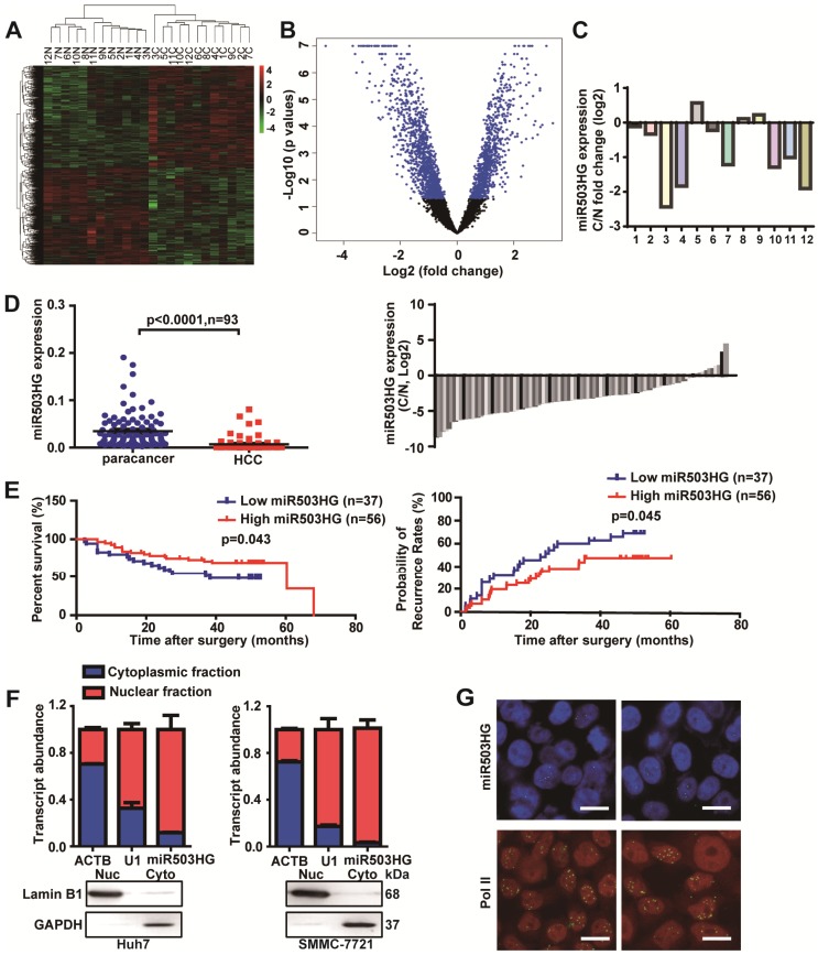
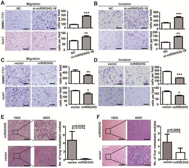
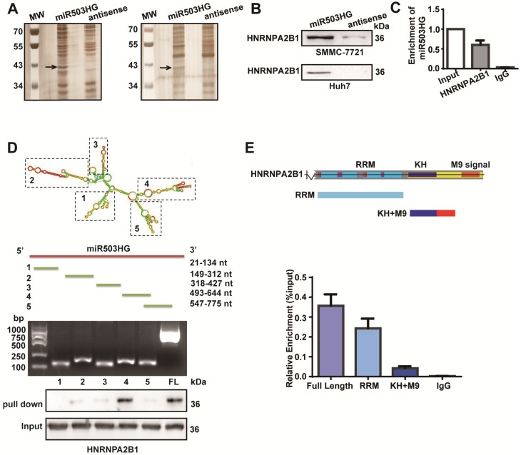
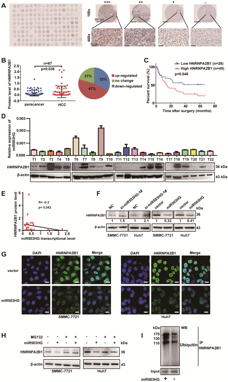
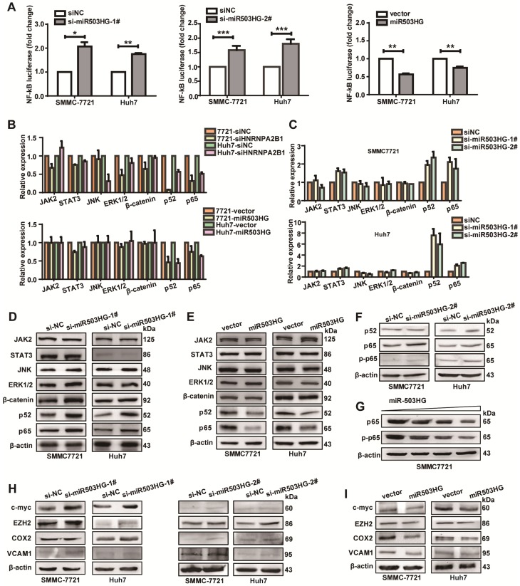
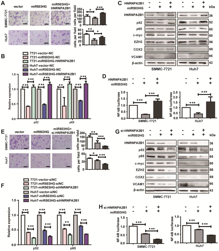
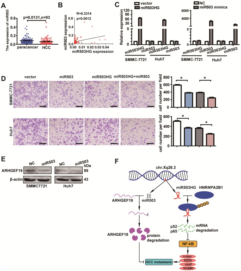
Long noncoding RNAs (lncRNAs) have been associated with hepatocellular carcinoma (HCC), but the underlying molecular mechanisms of their specific association with hepatocarcinogenesis have not been fully explored. Methods: miR503HG was identified by microarray and validated by real-time PCR. Survival analysis was evaluated using the Kaplan-Meier method and assessed using the log-rank test. In vitro and in vivo assays were preformed to explore the biological effects of miR503HG in HCC cells. The interaction of miR503HG with HNRNPA2B1 was identified by RNA pull-down and RNA immunoprecipitation. Expression of HNRNPA2B1 was examined by western blotting, immunofluorescence and immunohistochemical analyses, while HNRNPA2B1 ubiquitination was detected by immunoprecipitation. Results: We have identified 713 differentially expressed lncRNAs in 12 pairs of HCC tissues compared with corresponding noncancerous liver tissues. One of these lncRNAs, miR503HG, the host gene of miR503, is markedly decreased in HCC. Expression level of miR503HG is significantly associated with the time to recurrence and overall survival and is an independent risk factor for recurrence and survival. Enhanced expression of miR503HG could noticeably inhibit HCC invasion and metastasis in vitro and in vivo. Further investigation suggested that miR503HG could specifically interact with the heterogeneous nuclear ribonucleoprotein A2/B1 (HNRNPA2B1). miR503HG promoted HNRNPA2B1 degradation via the ubiquitin-proteasome pathway, which reduced the stability of p52 and p65 mRNA, and simultaneously suppressed the NF-κB signaling pathway in HCC cells. In addition, miR503HG can function synergistically with miR503 to inhibit HCC migration. Conclusion: Our findings support a role for miR503HG in tumor recurrence risk and survival prediction in HCC patients. We demonstrate a novel mechanism by which miR503HG inhibits the NF-κB signaling pathway and exerts its metastatic tumor suppression function through modulating the ubiquitination status of HNRNPA2B1.

Keywords: hepatocellular carcinoma; lncRNA; metastasis; miR503HG; prognosis.


Conflict of interest statement

Competing Interests: The authors have declared that no competing interest exists.

Figures

Figure 1
Figure 1
miR503HG is downregulated in HCC and is inversely associated with the patient prognosis. (A) Heatmap and (B) volcano plot illustrating the 713 differentially expressed lncRNAs between 12 pairs of cancerous tissues and corresponding adjacent non-cancerous liver tissues of HCC patients (fold change ≥ 1.5, P-value < 0.05). (C) The fold change of miR503HG expression in 12 HCC tissues. (D) Left: The expression of miR503HG in 93 pairs of HCC and noncancerous liver tissues. Right: the fold change of miR503HG in 93 HCC tissues. (E) Kaplan-Meier analysis of the correlation between miR503HG expression and overall survival or recurrence in 93 patients with HCC. Log-rank tests were used to determine statistical significance. (F) Real-time PCR analysis of the subcellular location of miR503HG in HCC cells. U1 snRNA (nuclear retained) and ACTB mRNAs (exported to cytoplasm) were used as controls. The values represent the median of 3 technical replicates. Data are the mean ± SD. (G) In situ hybridization analysis of the subcellular location of miR503HG in SMMC-7721 cells; Pol II enriched in the nucleus was used as a control. Scale bar = 20 μm.
Figure 2
Figure 2
miR503HG inhibits HCC cell invasion and metastasis in vitro and in vivo. (A-B) Migration assays of 5×10⁴ HCC cells or invasion assays of 1×10⁵ HCC cells with miR503HG knockdown by using siRNA-1#. Representative images are shown on the left, and the average number of cells per field at the indicated time points is shown on the right. Data are the mean ± SD. **P < 0.01 and ***P < 0.001. Scale bar = 100 μm. (C-D) Migration and invasion assays of SMCC-7721 and Huh7 cells that stably overexpress of miR503HG or control vector. Data are the mean ± SD. *P < 0.05, **P < 0.01 and ***P < 0.001. Scale bar = 100 μm. (E) Hematoxylin and eosin (H&E) staining of metastatic nodules in the lungs of nude mice 8 weeks after tail vein injection of Huh7 cells infected with lentivirus expressing miR503HG or the control. The numbers of metastatic nodules in the lungs of each mouse were counted and analyzed using Student's t-test. Data are presented as the mean ± SD (n = 9 mice per group). Scale bar = 200 μm (100x) or 50 μm (400x). (F) SMMC-7721 cells infected with lentivirus expressing miR503HG or the control vector were orthotopically injected into the livers of nude mice. 8 weeks later, the mice were sacrificed, and the livers were subjected to H&E staining. The numbers of liver metastatic nodules of each mouse were counted and analyzed using Students' t-test. Data are presented as the mean ± SD (n = 8 mice per group). Scale bar = 200 μm (100x) or 50 μm (400x).
Figure 3
Figure 3
miR503HG physically interacts with HNRNPA2B1 in HCC cells. (A) RNA pull-down assays were performed in SMMC-7721 cells twice. Biotinylated miR503HG (left lane) or antisense RNA (right lane) was incubated with cell extracts and targeted with streptavidin beads; the associated proteins were resolved on a gel. The highlighted region was submitted for mass spectrometry. (B) Western blotting analysis of the specific association of HNRNPA2B1 with miR503HG in SMMC-7721 and Huh7 cells. (C) RIP enrichment was determined as RNA associated with the HNRNPA2B1 IP relative to an input control. The data represent the average and standard deviation of three independent experiments. (D) Deletion mapping of the HNRNPA2B1-binding domain in miR503HG. Top: graphic illustration of predicted miR503HG secondary structure (LNCipedia, http://www.lncipedia.org), and the truncation of miR503HG according to the stem-loop structure. Middle: the in vitro transcribed full-length and deletion fragments of miR503HG. Bottom: Western blotting analysis of HNRNPA2B1 in protein samples pulled down by the different miR503HG constructs. (E) Deletion mapping of the miR503HG-binding domain in HNRNPA2B1. Top: diagrams of full-length HNRNPA2B1 and the domain-truncated fragments RNA recognition motif (RRM), hnRNP K homology domain (KH), M9 signal (MN). Bottom: qPCR analysis of HNRNPA2B1 retrieved by full-length or domain-truncated HNRNPA2B1-HA using a HA antibody. RIP assays were performed using Huh7 cells transfected with the indicated vectors.
Figure 4
Figure 4
miR503HG mediates ubiquitination and degradation of HNRNPA2B1 in HCC cells. (A) A tissue microarray with 80 pairs of HCC and corresponding noncancerous liver tissues was used to determine the protein expression level of HNRNPA2B1. (B) The protein level of HNRNPA2B1 was significantly increased in 47% of HCC tissues. (C) Kaplan-Meier analysis for OS was performed according to HNRNPA2B1 levels. (D) The transcriptional level of miR503HG and the protein expression level of HNRNPA2B1 in 22 HCC tissues. (E) Correlation analysis showed a negative relationship between miR503HG (x) and HNRNPA2B1 (y) in 22 HCC tissues (R = -0.2, P = 0.042). miR503HG and HNRNPA2B1 expression was normalized to β-actin. (F) HNRNPA2B1 protein expression in HCC cells with miR503HG knockdown or overexpression. (G) Immunofluorescence was used to compare the expression levels of HNRNPA2B1 between SMMC-7721 (or Huh7) cells with miR503HG overexpression and control cells. Scale bar = 20 μm. (H) SMMC-7721 and Huh7 cells expressing either miR503HG or control vector were cultured in the presence or absence of MG132 (20 μM) for 6 h. Cell lysates were then analyzed by western blotting. (I) Lysates from SMMC-7721 cells with miR503HG overexpression that were treated with MG132 were subjected to immunoprecipitation with anti-HNRNPA2B1 antibody followed by immunoblotting analysis with anti-ubiquitin or anti-HNRNPA2B1 antibody.
Figure 5
Figure 5
miR503HG inhibits the NF-κB signaling pathway in HCC cells. (A) Relative luciferase activity of NF-κB in miR503HG-downregulated or -upregulated SMMC-7721 and Huh7 cells. The values are presented as the mean ± standard error of the ratio of firefly luciferase activity to Renilla luciferase activity and are representative of three independent experiments. Data are the mean ± SD. *P < 0.05, **P < 0.01 and ***P < 0.001. (B) Real-time PCR analysis of the expression of JAK2, STAT3, JNK, ERK1/2, β-catenin, p52 and p65 in HNRNPA2B1 silenced (top) and miR503HG-overexpressing HCC cells (bottom). (C) The mRNA levels of JAK2, STAT3, JNK, ERK1/2, β-catenin, p52, and p65 in HCC cells with miR503HG knockdown. (D-E) Western blotting analysis shows changes in JAK2, STAT3, JNK, ERK1/2, β-catenin, p52, and p65 in miR503HG-downregulated and miR503HG-upregulated HCC cells. β-actin was used as the loading control. (F) Knockdown of miR503HG could increase the phosphorylation levels of p65 in HCC cells. (G) Overexpression of miR503HG decreased endogenous p65 and phospho-p65 in a dose-dependent manner. (H-I) The protein levels of the NF-κB downstream effectors were determined by western blotting analyses in miR503HG-downregulated or -upregulated HCC cells.
Figure 6
Figure 6
HNRNPA2B1 is required for miR503HG regulation of HCC cell migration and the NF-κB signaling pathway. (A) Migration assays of SMMC-7721 and Huh7 cells with overexpression of miR503HG and HNRNPA2B1. Scale bar = 100 μm. (B) The mRNA levels of p52 and p65 in SMMC-7721 and Huh7 cells that concomitantly overexpress miR503HG and HNRNPA2B1. (C) Western blotting analysis shows the changes of p52, p65 and the NF-κB downstream genes in HNRNPA2B1-restored HCC cells. (D) Relative luciferase activity of NF-κB in SMMC-7721 and Huh7 cells overexpressing HNRNPA2B1 with miR503HG. (E) Migration assays of HCC cells with miR503HG overexpression and HNRNPA2B1 knockdown. Scale bar = 100 μm. (F) The mRNA levels of p52 and p65 in miR503HG-overexpressing HCC cells with HNRNPA2B1 knockdown. (G) The protein levels of p52, p65 and the NF-κB downstream genes in miR503HG-overexpressing HCC cells with knockdown of HNRNPA2B1. (H) The NF-κB activity in HCC cells with miR503HG overexpression and HNRNPA2B1 knockdown. Data in (A-B, D-F, H) are the mean ± SD. *P < 0.05, **P < 0.01 and ***P < 0.001.
Figure 7
Figure 7
miR503HG functions synergistically with miR503 to suppress HCC cell migration and invasion. (A) Expression of miR503 in 93 pairs of HCC tissues and the corresponding normal liver tissues. U6B was used as the negative control. (B) The correlation of the expression levels of miR503 and miR503HG. Pearson's correlation was performed. (C) Expression of miR503 and miR503HG following overexpression of miR503HG and miR503 in SMMC-7721 and Huh7 cells. (D) Transwell migration assays of SMMC-7721 and Huh7 cells were performed after transient transfection of miR503 in miR503HG-upregulated or control HCC cells. Data are the mean ± SD. *P < 0.05. Scale bar = 100 μm. (E) Western blotting analysis shows changes in ARHGEF19 in miR503-upregulated SMMC-7721 and Huh7 cells. (F) A proposed model for illustrating the expression, function and mechanism of miR503HG in HCC metastasis.

References

    1. Thomas MB, Jaffe D, Choti MM, Belghiti J, Curley S, Fong Y, Gores G. et al. Hepatocellular carcinoma: consensus recommendations of the National Cancer Institute Clinical Trials Planning Meeting. J Clin Oncol. 2010;28:3994–4005. - PMC - PubMed
    1. Aldrighetti L, Pulitano C, Catena M, Arru M, Guzzetti E, Halliday J, Ferla G. Liver resection with portal vein thrombectomy for hepatocellular carcinoma with vascular invasion. Ann Surg Oncol. 2009;16:1254. - PubMed
    1. Ding J, Huang S, Wu S, Zhao Y, Liang L, Yan M, Ge C. et al. Gain of miR-151 on chromosome 8q24.3 facilitates tumour cell migration and spreading through downregulating RhoGDIA. Nat Cell Biol. 2010;12:390–99. - PubMed
    1. Yao J, Liang L, Huang S, Ding J, Tan N, Zhao Y, Yan M. et al. MicroRNA-30d promotes tumor invasion and metastasis by targeting Galphai2 in hepatocellular carcinoma. Hepatology. 2010;51:846–56. - PubMed
    1. Guo W, Qiu Z, Wang Z, Wang Q, Tan N, Chen T, Chen Z. et al. MiR-199a-5p is negatively associated with malignancies and regulates glycolysis and lactate production by targeting hexokinase 2 in liver cancer. Hepatology. 2015;62:1132–44. - PubMed

Publication types

MeSH terms

Substances