. 2018 Apr;8(3):291-300.
doi: 10.21037/qims.2018.03.04.

Spleen and splenic vascular involvement in acute pancreatitis: an MRI study

Affiliations

Spleen and splenic vascular involvement in acute pancreatitis: an MRI study

Chao-Lian Xie et al. Quant Imaging Med Surg. 2018 Apr.

Abstract

Background: To investigate the spleen and splenic vascular involvement in acute pancreatitis (AP) and their correlations with the severity of AP using magnetic resonance imaging (MRI).

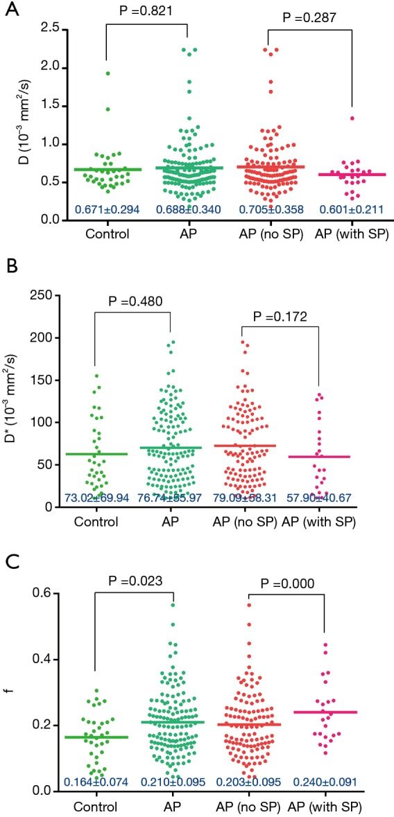
Methods: MRI of 239 patients with AP was retrospectively reviewed to assess splenic and splenic vascular complications, and the severity of AP. The severity of AP was graded by the MRI severity index (MRSI) and the New Revised Classification of AP 2012. The intravoxel incoherent motion (IVIM) parameters (D, D*, and f) of spleen were measured. Thirty-five subjects without pancreatic and splenic disorders were enrolled as controls for IVIM parameters.

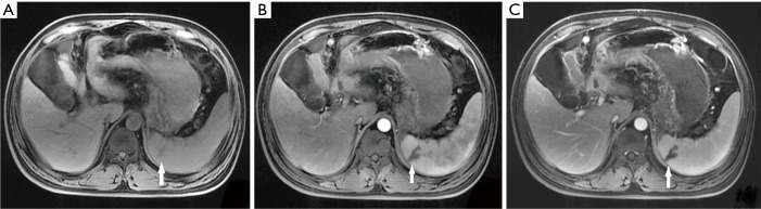
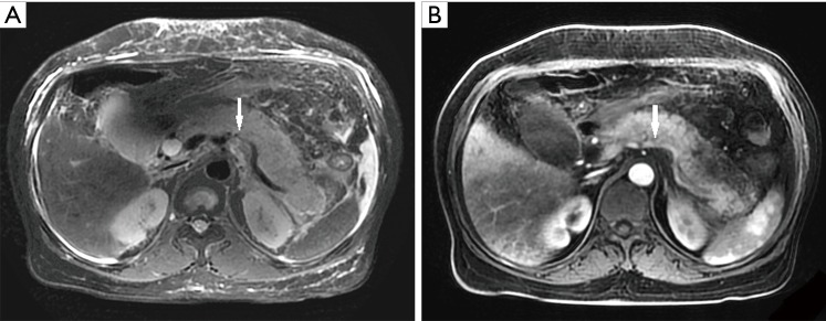
Results: Among the 239 patients with AP, splenomegaly (16.7%), splenic infarction (0.4%), splenic vein thrombosis (4.2%), phlebitis (7.5%) and arteritis (4.2%) were observed. Splenic vascular involvement was positively correlated with the severity of AP based on both the MRSI and the New Revised Classification of AP 2012 (P<0.05). In the control and AP groups, the splenic f values were (0.164±0.074) vs. (0.210±0.095) (P=0.023) respectively. In AP patients with and without splenomegaly, f = (0.240±0.091) vs. (0.203±0.095) (P<0.001).

Conclusions: Splenic vascular involvement and splenomegaly were common in AP. The vascular involvement was associated with the severity of AP. This complication should be considered when severity and prognosis of AP are assessed. Quantitative analysis of the spleen with IVIM might be a useful imaging biomarker for splenic perfusion changes in AP, especially in those with splenomegaly.

Keywords: Pancreas; acute pancreatitis (AP); magnetic resonance; spleen.


Conflict of interest statement

Conflicts of Interest: The authors have no conflicts of interest to declare.

Figures

Figure 1
Figure 1
A 45-year-old man with AP. The axial T1-weighted fat-suppressed image (A) shows a wedge of lower intensity in the spleen (arrows), which does not enhance in the enhanced arterial phase (B) and venous phase (C). AP, acute pancreatitis.
Figure 2
Figure 2
A 56-year-old woman with AP. The axial T2-weighted fat-suppressed image (A) shows a patchy high signal (arrow) in the splenic vein lumen that shows a filling defect (arrows) in the enhanced venous phase (B) and delayed phase (C). AP, acute pancreatitis.
Figure 3
Figure 3
A 38-year-old man with AP. The axial T2-weighted fat-suppressed image (A) shows the loss of the normal vascular flow void effect and a linear high signal of the splenic vein edge (arrow), which shows the poor enhancement of the vein and unsharpness of the splenic vein-edge (arrow) in enhanced venous phase (B). AP, acute pancreatitis.
Figure 4
Figure 4
A 45-year-old man with AP. The axial T2-weighted fat-suppressed image (A) shows the loss of the normal vascular flow void effect in the splenic artery (arrow) which shows the unsharpness of the splenic arterial edge (arrow) in enhanced arterial phase (B). AP, acute pancreatitis.
Figure 5
Figure 5
Scattered plots and mean of D, D*, and f of the control, AP, AP (no SP), and AP (with SP) patients. P value, analysis of variance. Mann-Whitney U-test is used to analyze the differences of splenic D, D*, and f between the control and AP group as well as the differences between the AP (no SP) and AP (with SP) group. AP, acute pancreatitis; SP, splenomegaly.

References

    1. Triester SL, Kowdley KV. Prognostic factors in acute pancreatitis. J Clin Gastroenterol 2002;34:167-76. 10.1097/00004836-200202000-00014 - DOI - PubMed
    1. Rau BM. Outcome determinants in acute pancreatitis. Am J Surg 2007;194:S39-44. 10.1016/j.amjsurg.2007.05.019 - DOI
    1. Xiping Z, Yan P, Xinmei H, Guanghua F, Meili M, Jie N, Fangjie Z. Effects of dexamethasone and Salvia miltiorrhizae on the small intestine and immune organs of rats with severe acute pancreatitis. Inflammation 2010;33:259-66. 10.1007/s10753-010-9180-9 - DOI - PubMed
    1. Shi C, Zhao X, Lagergren A, Sigvardsson M, Wang X, Andersson R. Immune status and inflammatory response differ locally and systemically in severe acute pancreatitis. Scand J Gastroenterol 2006;41:472-80. 10.1080/00365520500318965 - DOI - PubMed
    1. Xiping Z, Ruiping Z, Binyan Y, Li Z, Hanqing C, Wei Z, Rongchao Y, Jing Y, Wenqin Y, Jinjin B. Protecting effects of a large dose of dexamethasone on spleen injury of rats with severe acute pancreatitis. J Gastroenterol Hepatol 2010;25:302-8. 10.1111/j.1440-1746.2009.05999.x - DOI - PubMed

LinkOut - more resources