Histologic mimics of malignant melanoma
- PMID: 29774360
- PMCID: PMC6250758
- DOI: 10.11622/smedj.2018041
Histologic mimics of malignant melanoma
Abstract
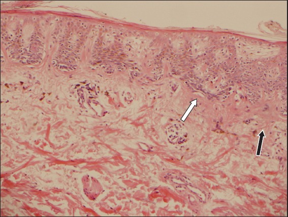
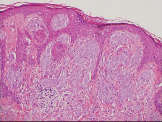
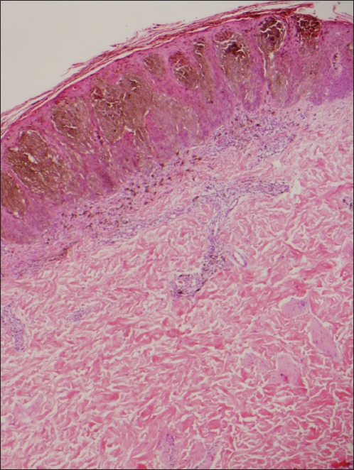
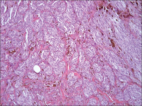
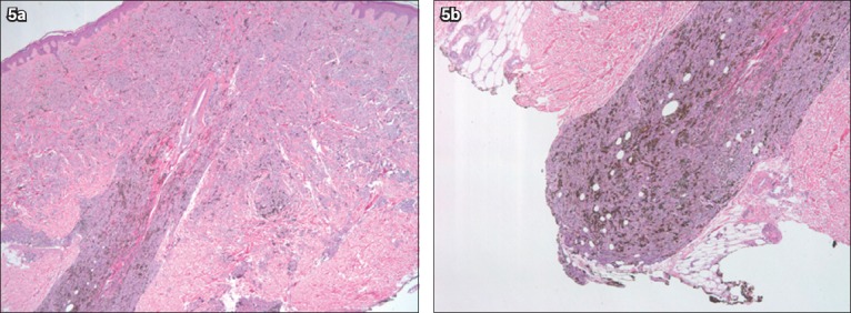
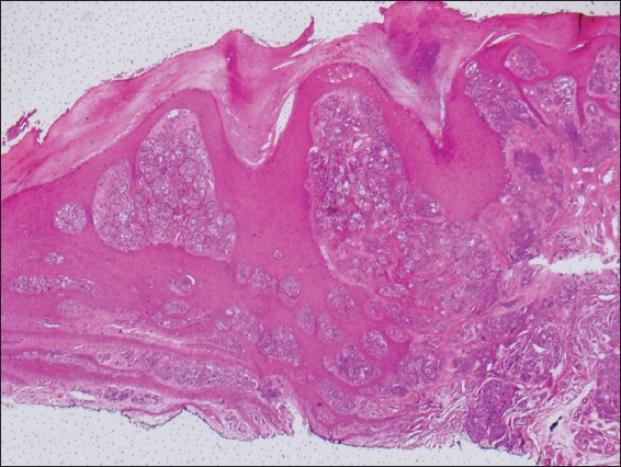
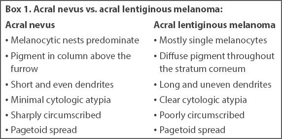
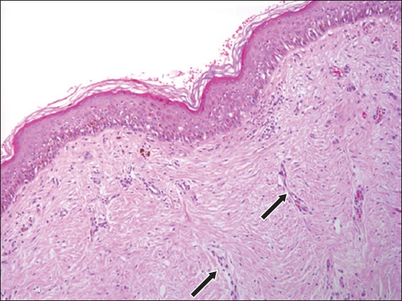
Although spongiotic (eczematous), psoriatic and cutaneous skin infections are among the most common in dermatology consultations, melanocytic lesions - including the different types of nevi and melanomas - are among those that cause a great deal of concern and stress to patients and their clinicians. A diagnosis of benign melanocytic nevus carries a very good prognosis. However, a diagnosis of melanoma might indicate more aggressive treatment, lifelong surveillance and a worse prognosis. Differentiating between these conditions is not always a straightforward process for clinicians and pathologists. Therefore, knowledge of melanoma mimickers is very important for clinicians in general, and dermatologists and pathologists in particular. In this review, we called attention to some of the more frequent benign but unusual melanocytic lesions that are of diagnostic concern for clinicians evaluating these cutaneous proliferations.
Keywords: deep penetrating nevus; dysplastic nevus; melanocytic lesions; melanoma; pigmented spindle cell nevus.
Copyright: © Singapore Medical Association.
Figures
References
-
- Rigel DS, Russak J, Friedman R. The evolution of melanoma diagnosis:25 years beyond the ABCDs. CA Cancer J Clin. 2010;60:301–16. - PubMed
-
- Weyers W. The ‘epidemic’of melanoma between under- and overdiagnosis. J Cutan Pathol. 2012;39:9–16. - PubMed
-
- Clarke LE. Dysplastic nevi. Clin Lab Med. 2011;31:255–65. - PubMed
-
- Kraemer KH, Greene MH, Tarone R, et al. Dysplastic naevi and cutaneous melanoma risk. Lancet. 1983;2:1076–7. - PubMed
Publication types
MeSH terms
LinkOut - more resources
Full Text Sources
Other Literature Sources
Medical