Review
. 2018 Aug;39(6):1107-1114.
doi: 10.1007/s00246-018-1906-8. Epub 2018 May 17.

Developmental Origin of the Cardiac Conduction System: Insight from Lineage Tracing

Affiliations
Review

Developmental Origin of the Cardiac Conduction System: Insight from Lineage Tracing

Rajiv A Mohan et al. Pediatr Cardiol. 2018 Aug.

Abstract

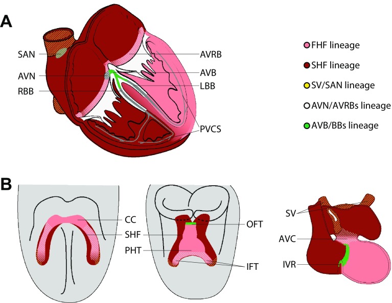
The components of the cardiac conduction system (CCS) generate and propagate the electrical impulse that initiates cardiac contraction. These interconnected components share properties, such as automaticity, that set them apart from the working myocardium of the atria and ventricles. A variety of tools and approaches have been used to define the CCS lineages. These include genetic labeling of cells expressing lineage markers and fate mapping of dye labeled cells, which we will discuss in this review. We conclude that there is not a single CCS lineage, but instead early cell fate decisions segregate the lineages of the CCS components while they remain interconnected. The latter is relevant for development of therapies for conduction system disease that focus on reprogramming cardiomyocytes or instruction of pluripotent stem cells.

Keywords: Cardiac conduction system; Cardiac development; Genetic inducible fate map; Lineage tracing.


Conflict of interest statement

Conflict of interest

The authors declare that they have no conflict of interest.

Ethical Approval

This article does not contain any studies with human participants or animals performed by any of the authors.

Figures

Fig. 1
Fig. 1
Developmental origin of the cardiac conduction system components. a The definitive cardiac conduction system (CCS, gray) consists of the sinoatrial node (SAN), the atrioventricular node (AVN) and ring bundles (AVRBs), the atrioventricular bundle (AVB), left and right bundle branches (LBB and RBB, respectively) and the peripheral ventricular conduction system (PVCS). The first and second heart field (FHF and SHF, respectively) contributions to the adult myocardium are visualized in pink (FHF-derived) and dark red (SHF-derived). b Mesodermal cells in the FHF differentiate and form the cardiac crescent (CC) at E7.5. Fusion of the cardiac crescent (E8) gives rise to the primary heart tube (PHT) and subsequently forms the left ventricle and part of the atrioventricular canal (AVC). Cardiogenic mesodermal cells in the SHF are continuously added to both poles of the heart tube and form the other myocardial components of the heart. The locations of the progenitor cells of the CCS components are depicted by yellow lines for the SV/SAN, blue lines for AVN/AVRBs and green dots for AVB/BBs. IFT inflow tract, IVR interventricular ring, OFT outflow tract, SV sinus venosus. Reproduced with permission from [3]
Fig. 2
Fig. 2
Lineage tracing identifies CCS progenitors and reveals its mode of development. a The definitive SAN includes a ‘head’ and ‘tail’ part, both expressing Tbx3. Tbx18 is specifically expressed in the ‘head’ and not in the ‘tail.’ However, using Tbx18-Cre;R26RLacZ/LacZ mice, the Tbx18+ progenitors were found to contribute to both parts of the SAN. b Spatio-temporal expression pattern of Gja5 at several stages. At E10.5 Gja5 is expressed transmurally in the embryonic left ventricular myocardium. At E14.5, expression is downregulated in the compact myocardium of the left ventricle and maintained within the trabeculae. At the adult stage, Gja5 is only expressed in the PVCS of the left ventricle. c Labeling E10.5 embryonic Gja5+ cardiomyocytes using Tamoxifen in an inducible Cre model (Gja5-CreERT2-IRESmRFP;R26R-YFP) shows a contribution to the formed PVCS and ventricular working myocardium demonstrating a common origin from Gja5+ cells and downregulation of Gja5 in the compact myocardium. Tbx18 lineage tracing is adapted from [17], Gja5 expression pattern from [23], and the Gja5 lineage tracing are unpublished results from [24]

References

    1. Wolf CM, Berul CI. Inherited conduction system abnormalities—One group of diseases, many genes. J Cardiovasc Electrophysiol. 2006;17:446–455. doi: 10.1111/j.1540-8167.2006.00427.x. - DOI - PubMed
    1. Mangoni ME, Nargeot J. Genesis and regulation of the heart automaticity. Physiol Rev. 2008;88:919–982. doi: 10.1152/physrev.00018.2007. - DOI - PubMed
    1. Mohan R, Boukens BJ, Christoffels VM. Lineages of the Cardiac Conduction System. J Cardiovasc Dev Dis. 2017;4:5. doi: 10.3390/jcdd4020005. - DOI - PMC - PubMed
    1. Hariman RJ, Hoffman BF, Naylor RE. Electrical activity from the sinus node region in conscious dogs. Circ Res. 1980;47:775–791. doi: 10.1161/01.RES.47.5.775. - DOI - PubMed
    1. Draper MH, Weidmann S. Cardiac resting and action potentials recorded with an intracellular electrode. J Physiol. 1951;115:74–94. doi: 10.1113/jphysiol.1951.sp004653. - DOI - PMC - PubMed