Review
. 2018 Dec;596(23):5723-5756.
doi: 10.1113/JP275376. Epub 2018 Jul 15.

Physiology and molecular biology of barrier mechanisms in the fetal and neonatal brain

Affiliations
Review

Physiology and molecular biology of barrier mechanisms in the fetal and neonatal brain

Norman R Saunders et al. J Physiol. 2018 Dec.

Abstract

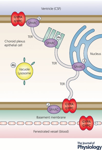
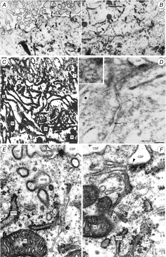
Properties of the local internal environment of the adult brain are tightly controlled providing a stable milieu essential for its normal function. The mechanisms involved in this complex control are structural, molecular and physiological (influx and efflux transporters) frequently referred to as the 'blood-brain barrier'. These mechanisms include regulation of ion levels in brain interstitial fluid essential for normal neuronal function, supply of nutrients, removal of metabolic products, and prevention of entry or elimination of toxic agents. A key feature is cerebrospinal fluid secretion and turnover. This is much less during development, allowing greater accumulation of permeating molecules. The overall effect of these mechanisms is to tightly control the exchange of molecules into and out of the brain. This review presents experimental evidence currently available on the status of these mechanisms in developing brain. It has been frequently stated for over nearly a century that the blood-brain barrier is not present or at least is functionally deficient in the embryo, fetus and newborn. We suggest the alternative hypothesis that the barrier mechanisms in developing brain are likely to be appropriately matched to each stage of its development. The contributions of different barrier mechanisms, such as changes in constituents of cerebrospinal fluid in relation to specific features of brain development, for example neurogenesis, are only beginning to be studied. The evidence on this previously neglected aspect of brain barrier function is outlined. We also suggest future directions this field could follow with special emphasis on potential applications in a clinical setting.

Keywords: amino acids; blood vessels; blood-brain barrier; cerebrospinal fluid; choroid plexus; electrolytes; electron microscope; embryo; endothelium; epithelium; gene transcripts; immunohistochemistry; ion gradients; meninges; protein; tight junctions; transport.


Figures

Figure 1
Figure 1. Schematic diagram (middle right) of the five main barrier interfaces (A–D and F) in the adult and developing brain and an additional one present only in the embryo (E)
The barrier‐forming cellular layers at each interface are coloured green. A, the blood–brain barrier is situated at the level of cerebral blood vessels (BV). Tight junctions (tj, arrowhead) are present between the endothelial cells (EC) restricting the paracellular cleft. AE, end feet from astroglial cells; bm, basement membrane; PC, pericytes. Other important components of this interface are a basement membrane of extracellular matrix, within which are embedded pericytes (Engelhardt & Sorokin, 2009) surrounding the endothelial cells (Daneman et al. 2010b, Errede et al. 2014). Astroglial end feet encircle cerebral blood vessels during the first 2–3 weeks of postnatal development in rodents (Caley & Maxwell, 1970) although the encirclement appears to be less complete than previously thought (Korogod et al. 2015). The contribution of astrocytes to development and maintenance of barrier properties is controversial as will be discussed. These cellular structures are known collectively as the neurovascular unit (Neuwelt, 2004). B, the blood–CSF barrier is situated in the choroid plexus within each brain ventricle. Barrier‐forming cells are the epithelial cells (CPE), which have tight junctions at their apical side (CSF facing, arrowheads). Blood vessels (BV) are fenestrated and do not form a barrier (arrows); apical microvilli increase exchange surface of epithelial cells to the internal CSF (i‐CSF) C, circumventricular organs (including median eminence, pineal gland, area postrema, subfornical organ). Blood vessels have permeability characteristics similar to elsewhere in the body and have the functional property of allowing feedback penetration of peptide hormones controlled by the hypothalamic–pituitary axis. These peptides and other molecules are prevented from entering the CSF by tanycytes (TC), the specialized ependymal cells of these brain areas, connected by tight junctions between their apices (arrowhead); entry into the rest of the brain is prevented by tight junctions between astroglial cells (GC). Away from the tanycyte layer, ependymal cells lining the ventricular system are linked by gap junctions that allow free exchange between the CSF and brain interstitial fluid. D, ependyma in adult brain. Apart from areas where there are specialized tanycytes, ependymal cells are linked by gap junctions that do not restrict exchange of even large molecules, such as proteins, between CSF and interstitial space of brain (arrows). E, the embryonic CSF–brain barrier. In early brain development, strap junctions (arrowheads) are present between adjacent neuroepithelial cells (NE); these form a barrier restricting the movement of larger molecules, such as proteins, but not smaller molecules. F, the meningeal barrier is structurally the most complex of all the brain barriers. Barrier‐forming cells are the outer layer of the arachnoid membrane (the arachnoid barrier cells; ABC); these have tight junctions (arrowheads) between adjacent cells forming a barrier between the outer cerebrospinal fluid (o‐CSF) in the subarachnoid space (SAS) and more superficial dural layers (dural border cells (DBC) and the dura mater). Blood vessels (BV) in the SAS have tight junctions with similar barrier characteristics as cerebral blood vessels without surrounding pericytes and astrocytic end‐feet. Blood vessels within the dura mater are fenestrated (f‐BV). bm, basement membrane; gl, glia limitans. Redrawn from Saunders et al. (2016b).
Figure 2
Figure 2. Morphology of the brain barriers illustrated in Fig. 1
Aa, the blood–brain barrier. Light micrograph showing the localization of biotin ethylenediamine (BED) in the neocortex of opossum at P5 20–25 min after an intraperitoneal injection. Note that the staining for BED is most visible within the vessels, in the marginal and subplate zones. cp, cortical plate; mz, marginal zone; sp, subplate. Ab, localization of biotin–dextran (BDA3000) in the neocortex of P2 opossum 20–25 min after an intraperitoneal injection. Arrowhead points to site of the tight junction. Note that the marker is prevented from passing through the intercellular space by the tight junction. From Ek et al. (2006). B, the blood–CSF barrier. Lateral ventricular choroid plexus (blood–CSF barrier) in P13 Monodelphis. Tight junction excludes entry of BDA3000 into CSF. Labelled dextran present in one large and several smaller endosomes. From Ek et al. (2003). C, Circumventricular organs. a, adjacent tanycytes facing the CSF in circumventricular organs are connected by an extensive network of apical tight junctional strands shown here by freeze fracture of the adult Mongolian gerbil subcommissural organ. b, a thin section electron micrograph of the same region in a neonatal animal shows multiple ‘kissing points’ (arrows) between neighbouring tanycytes indicating a complete occlusion of the paracellular pathway. From Madsen & Møllgård (1979). D, ependyma in adult brain. Junctional configuration in the ependymal layer of 125‐day sheep fetus similar to those found in mature ependyma. ZA, zonulae adherens. Note unobstructed intercellular space. E, the embryonic CSF–brain barrier. a, thin‐section electron micrograph of the neuroepithelial lining of the cerebral vesicle from an E19 sheep. The junctional zone exhibits very narrow intercellular clefts which at places seem to be totally occluded (arrows). This junctional configuration has been named ‘strap’ junction. b, characteristic freeze fracture single strand of strap junction perpendicular to CSF surface. From Møllgård et al. (1987). F, the meningeal barrier. Distribution of claudin‐11 immunoreactivity in sagittal sections of E18 rat (a and b) and 21st wpc human (c) brain. Demonstrates a strong reactivity of the entire arachnoid barrier cell layer = arachnoid blood–CSF barrier (aB‐CSFB, arrowheads). From Brøchner et al. (2015). CM, cisterna magna; EFL, radial glial end feet layer; SAS, subarachnoid space; TC, tentorium cerebelli.
Figure 3
Figure 3. Ion gradients between CSF and plasma in developing and adult rat brain
A characteristic of CSF is its stable ionic composition that differs from that of plasma to an extent that cannot be explained by ultrafiltration, as was once thought. Data for CSF and plasma (mEq L−1 H2O) are from Amtorp & Sørensen (1974) and for intracellular ions (mmol L−1 H2O) from Fig. 8 in Johanson & Murphy (1990). The gradients are the consequence of the complex interactions between enzymes (notably carbonic anhydrase) ion transporters and ion channels, as illustrated in Fig. 4. The CSF secretion rate in the embryo and newborn is much lower than in the adult (Bass & Lundborg, 1973; Johanson & Woodbury, 1974), which is perhaps explained by the much lower expression of carbonic anhydrase and ATPases in the developing choroid plexus, as indicated in Fig. 4. Redrawn from Saunders et al. (2016b).
Figure 4
Figure 4. Localization of proteins for ion transporters, channels and associated enzymes and identification of their corresponding genes in adult and immature rat choroid plexus
CSF secretion results from coordinated intracellular carbonic anhydrase activity and transport of ions and water from basolateral membrane to cytoplasm, then sequentially across apical membrane into the cerebral ventricles (Davson & Segal, 1996; Speake et al. 2001; Praetorius & Damkier, 2017). This process has only been studied in adult choroid plexus (Brown et al. 2004; Praetorius & Damkier, 2017). The membrane and intracellular locations of the ion channels, transporters and enzymes indicated are from Praetorius & Damkier (2017). Data from Liddelow et al. (2013) compares expression of these genes and other functionally related genes in E15 and adult rat lateral ventricular choroid plexus. Blue indicates the genes that are upregulated (enriched) in the adult. Light red indicates genes that are expressed at a higher level at E15. We have assumed the same cellular/membrane location for members of the same gene family. The genes all had substantial but variable transcript numbers in the RNA‐Seq analysis. In some cases where a gene was upregulated in the adult, the transcript number was also high in the embryo, suggesting this transporter or channel was likely to be functionally effective at both ages, e.g. the K+ channel Kcnj13 (Kir7.1), Slc12a2 (NKCC1) a Na+–K+–Cl exchanger and Slc4a2 (NBCe2) a coupled Na+–HCO3 pump. ATPB1 (Atpb1b1) is a Na+/K+‐ATPase. Green indicates genes that were expressed at similar levels at the two ages. There are many more channels that show age‐related differential expression in choroid plexus, the functions of which are unclear. Redrawn from Liddelow et al. (2016) with additional data from Liddelow et al. (2013).
Figure 5
Figure 5. Influx transporters at the blood–brain barrier
These are mainly SLC (solute carrier) transporters. See Hediger (2013) for comprehensive review. Only transporters for which there is physiological evidence for function are listed. As indicated in Table 2, transcripts for many more Slcs have been identified in molecular screens. Many of these genes are found in both endothelial cells of the blood–brain barrier and epithelial cells of the choroid plexuses. Others are unique to each interface as summarized in Table 2. Note, many metal ions that are potentially toxic can be carried in via some of these transporters.
Figure 6
Figure 6. Efflux transporters at the blood–brain barrier
These are mainly ATP‐binding cassette (ABC) transporters. Some, e.g. P‐glycoprotein (PGP; ABCB1), reduce entry into cells. Others, e.g. multidrug resistance protein (MRPs; ABCCs), ligand (drug or toxin) combines with glutathione, glucuronic acid or sulphate in cells before efflux. BCRP, breast cancer resistance protein (ABCG2). ‘Others’ include SLC efflux (SLCO) transporters.
Figure 7
Figure 7. Total protein concentration in CSF of various species at different post‐conceptional ages
Ordinate: total protein concentration mg (100 mL CSF)−1. Abscissa: post‐conceptional age in days. Filled circles indicate time of birth. Data for sheep from Dziegielewska et al. (1980a); pig (Cavanagh et al. 1982); rat (Dziegielewska et al. 1981; Checiu et al. 1984); tammar wallaby, Macropus eugenii (Dziegielewska et al. 1986); opossum, Monodelphis domestica (Dziegielewska et al. 1989); chick, (Birge et al. 1974); rabbit (Ramey & Birge (1979). Adult values not shown. For mammalian species, including human (see Davson, 1967) and marsupials mean values are between 23 and 31 mg (100 mL)−1. Chicken is 141 mg (100 mL)−1.
Figure 8
Figure 8. Penetration of human plasma proteins from blood into CSF of 60‐day sheep fetuses
Human plasma was injected intravenously and blood was sampled to give an estimate of mean plasma concentration. At the times indicated CSF was sampled from cisterna magna. Concentrations of individual proteins in CSF and plasma were estimated by radial immunodiffusion assay. Abscissa: time in hours following i.v. injection; ordinate: CSF concentration/plasma concentration × 100. Steady state indicates CSF/plasma ratio for naturally occurring sheep proteins. Mean ± SEM for three to six experiments. All injected proteins were human (h‐) except for s‐Alb (35S‐sheep albumin) g‐Alb (goat albumin) and b‐Alb (bovine albumin) measured using sheep anti‐goat or anti‐bovine albumin antiserum). Experimental details and data are from Dziegielewska et al. (1980a,1980b, 1991). α1AT, α1‐antitrypsin; AFP, α‐fetoprotein; Alb, albumin; IgG, immunoglobulins; Trf, transferrin. The sheep fetus does not possess any IgG of its own, and hence no steady state ratio is shown. Note (i) there is an apparent relation between molecular size and permeability (the largest molecule, IgG, has the lowest ratio and the smallest molecule, AFP, has the largest ratio; however, all of these ratios except for IgG are higher than would be expected from passive diffusion (Saunders, 1992); and (ii) albumin from different species may have different ratios, which suggests that there is a selective mechanism that transports proteins from plasma to CSF. The route of protein transfer appears to be via the epithelial cells of the choroid plexus (Jacobsen et al. 1983; Dziegielewska et al. 1991).
Figure 9
Figure 9. Cellular distribution of mouse albumin–SPARC (A–C) and human albumin–SPARC (D–F)
Demonstrated by in situ Proximity Ligation Assay (in situ PLA) signals in the lateral ventricular choroid plexus at P2 (A and D), P10 (B and E) and adult (C and F). Note at P2 that most of the signal was distributed within blood vessels (BV), often associated with red blood cells. Under this magnification it is possible to distinguish positive signals distributed in the basolateral cytoplasm of choroid plexus epithelial cells (arrows). In contrast to the mouse albumin–SPARC signal, the human albumin–SPARC signal (D) was very rarely found and nearly always only associated with blood vessels (BV). Only one positive signal was found and it appears to be located in the extended extracellular space (arrowhead). At P10 (B and E) a very strong signal was visible for mouse albumin–SPARC (B) in many plexus cells distributed throughout the whole cytoplasm, blood vessels (BV) and also in the CSF. The human albumin–SPARC signal (E) was generally only present in blood vessels (BV) but a very occasional signal was detected in the apparent extended extracellular space (arrowhead). The CSF space was negative. In the adult (C and F) a mouse albumin–SPARC signal was distributed clearly throughout the cytoplasm of some choroid plexus epithelial cells (one cell marked with an asterisk). The positive signal was also detected in the ependymal (EP) and subependymal layers of the brain. The human albumin–SPARC signal (F) was visible in blood vessels (BV) but not in the CSF and only very sporadically in the plexus epithelium (two positive red dots are indicated by arrows). Otherwise plexus epithelial cells (boxes) showed no in situ PLA signal. BV, blood vessels; CSF, cerebrospinal fluid; EP, ependymal. Same magnification, scale bar is 20 μm. From Liddelow et al. (2014).
Figure 10
Figure 10. Proposed transepithelial pathway for albumin through choroid plexus epithelial cells
Cartoon of suggested routes of albumin transfer from plasma into CSF across the choroid plexus epithelium. GYPA/C in the endothelial cells may deliver albumin to the basement membrane (1) from where it can be taken up into the plexus epithelium by GYPA/C or SPARC (2). From here albumin may travel along a SPARC‐specific pathway through the tubulocisternal endoplasmic reticulum (3) (see Fig. 11) and Golgi (4a), or via a VAMP‐mediated pathway in vacuoles, lysosomes or multivesicular bodies (4b). On the apical surface of the plexus epithelium, GYPA/C may be involved in efflux of protein from the cell into the CSF of the ventricles (5), as validated by extensive GYPA immunoreactivity in embryonic plexus (Liddelow et al. 2012). In the adult, the lack of immunoreactivity in the endoplasmic reticulum and Golgi (Liddelow et al. 2012) along with increased expression of gene products for VAMP molecules suggests that the majority of transport possibly occurs via VAMP‐mediated vesicular/lysosomal transport such as shown in (4b). CSF, cerebrospinal fluid; GYPA, glycophorin A; GYPC, glycophorin C; SPARC, secreted protein acidic and rich in cysteine; VAMP, vesicle‐associated membrane proteins. Redrawn from Liddelow et al. (2012).
Figure 11
Figure 11. Tubulocisternal endoplasmic reticulum (TER) in fetal sheep choroid plexus epithelial cells
A and B, electron micrographs of E60 fetal sheep choroid plexus. Alcian blue in Krebs solution was injected i.v. 10 min prior to fixation. Alcian blue is electron dense and binds to plasma albumin. Particulate precipitate (P in A) is visible within tubular endoplasmic reticulum (TER; dark arrows in B), which extends close to the lateral cell membrane (LM). JC, junctional complex separating the lateral intercellular space from lateral ventricular CSF. Curved arrows in A indicate precipitated Alcian blue on apical cell membrane; open arrows indicate close contact of TER with apical cell membrane, exposed to CSF. From Møllgård & Saunders (1975). C, high voltage double impregnation thick section EM of E60 fetal sheep choroid plexus. Note the extensive network of TER with contacts to CSF surface (uppermost in micrograph) and close association of TER with Golgi complex (G) and mitochondria (M). From Møllgård & Saunders (1977). D, electron micrograph of ultracryosection from E60 fetal sheep choroid plexus immunolabelled for human (HA) 6 nm gold particles and sheep albumin (SA) 12 nm gold particles (arrows). Gold particles labelling each of the albumins are colocalized within the same TER‐cistern (star) and a multivesicular body (MVB; inset). Scale bar, 0.2 μm. From Balslev et al. (1997a). E and F, fetal sheep choroid plexus (E60) double impregnation technique. Profiles of rough endoplasmic reticulum (RER) and TER system. Note TER termination on apical plasma membrane via a caveola (arrowhead in F). MVB, multivesicular body; M, mitochondrion. From Møllgård & Saunders (1977).

References

    1. Abbott NJ (2002). Astrocyte‐endothelial interactions and blood‐brain barrier permeability. J Anat 200, 629–638. - PMC - PubMed
    1. Abbott NJ, Patabendige AA, Dolman DE, Yusof SR & Begley DJ (2010). Structure and function of the blood‐brain barrier. Neurobiol Dis 37, 13–25. - PubMed
    1. Adinolfi M (1985). The development of the human blood‐CSF‐brain barrier. Dev Med Child Neurol 27, 532–537. - PubMed
    1. Adinolfi M & Haddad SA (1977). Levels of plasma proteins in human and rat fetal CSF and the development of the blood‐CSF barrier. Neuropadiatrie 8, 345–353. - PubMed
    1. Akiyama T, Kobayashi K, Higashikage A, Sato J & Yoshinaga H (2014). CSF/plasma ratios of amino acids: reference data and transports in children. Brain Dev 36, 3–9. - PubMed

Substances