. 2018 Aug;596(16):3675-3693.
doi: 10.1113/JP275067. Epub 2018 Jul 11.

Diabetes-induced microvascular complications at the level of the spinal cord: a contributing factor in diabetic neuropathic pain

Affiliations

Diabetes-induced microvascular complications at the level of the spinal cord: a contributing factor in diabetic neuropathic pain

N Ved et al. J Physiol. 2018 Aug.

Abstract

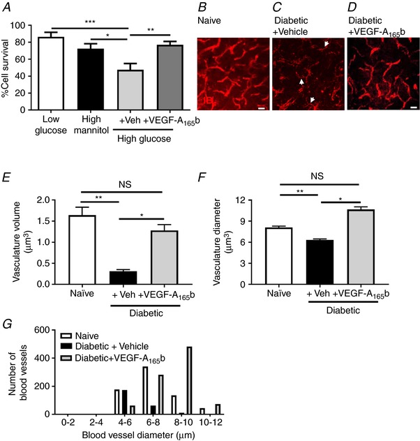
Key points: Diabetes is thought to induce neuropathic pain through activation of dorsal horn sensory neurons in the spinal cord. Here we explore the impact of hyperglycaemia on the blood supply supporting the spinal cord and chronic pain development. In streptozotocin-induced diabetic rats, neuropathic pain is accompanied by a decline in microvascular integrity in the dorsal horn. Hyperglycaemia-induced degeneration of the endothelium in the dorsal horn was associated with a loss in vascular endothelial growth factor (VEGF)-A165 b expression. VEGF-A165 b treatment prevented diabetic neuropathic pain and degeneration of the endothelium in the spinal cord. Using an endothelial-specific VEGFR2 knockout transgenic mouse model, the loss of endothelial VEGFR2 signalling led to a decline in vascular integrity in the dorsal horn and the development of hyperalgesia in VEGFR2 knockout mice. This highlights that vascular degeneration in the spinal cord could be a previously unidentified factor in the development of diabetic neuropathic pain.

Abstract: Abnormalities of neurovascular interactions within the CNS of diabetic patients is associated with the onset of many neurological disease states. However, to date, the link between the neurovascular network within the spinal cord and regulation of nociception has not been investigated despite neuropathic pain being common in diabetes. We hypothesised that hyperglycaemia-induced endothelial degeneration in the spinal cord, due to suppression of vascular endothelial growth factor (VEGF)-A/VEGFR2 signalling, induces diabetic neuropathic pain. Nociceptive pain behaviour was investigated in a chemically induced model of type 1 diabetes (streptozotocin induced, insulin supplemented; either vehicle or VEGF-A165 b treated) and an inducible endothelial knockdown of VEGFR2 (tamoxifen induced). Diabetic animals developed mechanical allodynia and heat hyperalgesia. This was associated with a reduction in the number of blood vessels and reduction in Evans blue extravasation in the lumbar spinal cord of diabetic animals versus age-matched controls. Endothelial markers occludin, CD31 and VE-cadherin were downregulated in the spinal cord of the diabetic group versus controls, and there was a concurrent reduction of VEGF-A165 b expression. In diabetic animals, VEGF-A165 b treatment (biweekly i.p., 20 ng g-1 ) restored normal Evans blue extravasation and prevented vascular degeneration, diabetes-induced central neuron activation and neuropathic pain. Inducible knockdown of VEGFR2 (tamoxifen treated Tie2CreERT2 -vegfr2flfl mice) led to a reduction in blood vessel network volume in the lumbar spinal cord and development of heat hyperalgesia. These findings indicate that hyperglycaemia leads to a reduction in the VEGF-A/VEGFR2 signalling cascade, resulting in endothelial dysfunction in the spinal cord, which could be an undiscovered contributing factor to diabetic neuropathic pain.

Keywords: diabetes; endothelial; pain.


Figures

Figure 1
Figure 1. Diabetes‐induced neuropathic pain is associated with a reduction in spinal cord vasculature and a decrease in VEGF‐A165b expression
A, diabetes resulted in a reduction in mechanical withdrawal threshold measured by pincher, when compared with naïve age‐matched animals (* P < 0.05, n = 5 per group). B, blood vessels identified (IB4) in the deeper laminar layers of the spinal cord (layers III–VI) of the naïve animal (C) with a decline in vascular staining in the diabetic animal. D, there was a reduction in blood vessel (CD31/IB4+ve) number (* P <0.05, n = 4 per group) [E = naïve, F = diabetic; reduced diameter (* P < 0.05, n = 4 per group)] as well as diameter (G) in the lumbar spinal cord in diabetic animals compared with naïve controls. H, immunoblot of pan‐VEGF, VEGF‐A165b and actin in lysates from spinal cord of normal and diabetic animals. I, densitometry analysis demonstrates no change in pan‐VEGF‐A expression and a decrease in VEGF‐A165b expression in diabetic lumbar spinal cord versus naïve animals (* P < 0.05, n = 5 per group). Scale bars: B and C = 40 μm, E and F= 20 μm. [Color figure can be viewed at http://wileyonlinelibrary.com]
Figure 2
Figure 2. Diabetes‐induced vascular impairment in the spinal cord
A, isolated spinal cord endothelial cells demonstrated increased cell death in 50 mm glucose when compared with 50 mm mannitol and 5 mm glucose (* P < 0.05, *** P < 0.001). VEGF‐A165b treatment prevented high glucose‐induced endothelial cell death (** P < 0.01). B, IB4 stained vasculature in the spinal cord of naïve age matched controls was compared with that in (C) diabetic + vehicle (arrowheads = vessels smaller than 6 μm) and (D) diabetic + VEGFA165b. E, there was a significant reduction in total volume of the microvasculature in the spinal cord of the diabetic + vehicle group in addition to (F) a reduction in vessel diameter compared with naïve controls (** P < 0.01, *** < 0.001, n = 4 per group). E and F, VEGF‐A165b treatment prevented the diabetes‐induced vascular degeneration in the lumbar spinal cord (** P < 0.01, n = 4 per group). G, VEGF‐A165b treatment also prevents the diabetes‐induced decrease in larger and intermediate microvessels. Scale bar: B–D = 25 μm. [Color figure can be viewed at http://wileyonlinelibrary.com]
Figure 3
Figure 3. Diabetes‐induced degeneration of the endothelium
Immunoblots using dual colour far red imaging for endothelial markers (A, VE‐cadherin, CD31; and B, occludin and actin) demonstrated (C) a non‐significant reduction in VE‐cadherin expression and significant reductions in (D) CD31 and (E) occludin in the diabetic + vehicle group compared with naïve and diabetic + VEGF‐A165b animals (* P < 0.05, n = 5).
Figure 4
Figure 4. Reduced vascular functionality in the spinal cord of diabetic rats
There was a significant reduction in Evans blue solute flux in the lumbar spinal cord of diabetic animals after (A) 1 week (naïve, n = 9; diabetes, n = 12; *** P < 0.001) and (B) 8 weeks (* P < 0.05, n = 4/5 per group). VEGF‐A165b treatment prevented the diabetes‐induced reduction in solute flux within the lumbar spinal cord at 8 weeks (* P < 0.05, n = 4/5 per group). C, there was no change in solute flux in the brain of any treatment group (naïve vs. diabetic + vehicle: P = 0.52; diabetic + vehicle vs. diabetic + VEGF‐A165b: P ≥ 0.99; n = 4/5 per group).
Figure 5
Figure 5. Diabetes‐induced dysfunction of microvasculature in the spinal cord and neuropathic pain is reversed by VEGF‐A165b
A, diabetic + vehicle animals demonstrated a decrease in mechanical withdrawal threshold (vF hairs) and (B) reduced withdrawal latency to heat compared with both naïve and diabetic + VEGF‐A165b treated groups (** P < 0.01, *** P < 0.001 naïve vs. diabetic + vehicle; # < 0.001 diabetic + VEGF‐A165b vs. diabetic + vehicle, n = 5 per group). Arrow highlights onset of VEGF‐A165b treatment. C, cleaved caspase‐3 (red = CC3) and sensory neuron (green = NeuN) staining in the spinal cord (scale bar = 40 μm). D, there was an increase in CC3 expression in sensory neurons in the superficial lamina (I and II) of the dorsal horn of the spinal cord in the diabetic + vehicle groups compared with naïve age‐matched controls and VEGF‐A165b treated diabetic animals. E, there was an increased number of CC3‐positive dorsal horn sensory neurons (arrows) in the dorsal horn of the spinal cord in the diabetic + vehicle groups compared with naïve age‐matched controls and VEGF‐A165b treated diabetic animals. This is graphically represented in (F) (* P < 0.05, n = 4). [Color figure can be viewed at http://wileyonlinelibrary.com]
Figure 6
Figure 6. Diabetes‐induced hyperactivity in sensory neurons of the dorsal horn in the spinal cord
A and B, immunoreactivity of a marker of central sensitisation, c‐fos (red; NeuN green), was increased in sensory neurons in the dorsal horn of the spinal cord in diabetic + vehicle animals versus naïve animals. This was reduced by VEGF‐A165b treatment. C, there was an increase of c‐fos expression in sensory neurons in all lamina of the dorsal horn (I–V) in the diabetic + vehicle group when compared to naïve and diabetic + VEGF‐A165b groups (* P < 0.05, ** P < 0.001, *** P < 0.001; n = 4). [Color figure can be viewed at http://wileyonlinelibrary.com]
Figure 7
Figure 7. Tamoxifen induced VEGFR2 knockout
VEGFR2 was inducibly knocked out in endothelial cells by crossing vegfr2 fl/fl with Tie2CreERT2 mice and treating with tamoxifen (VEGFR2ECKO). A, in lung tissue, VEGFR2 protein was detected in tamoxifen‐treated vegfr2 fl/fl mice lacking Cre (CTL), but not in VEGFR2ECKO mice. B, immunoblot densitometry demonstrated reduced VEGFR2 protein in the VEGFR2ECKO mice compared with controls (* P<0.05, n = 6 per group). C and D, VE‐cadherin expression was also reduced in lung tissue of the VEGFR2ECKO mice (* P < 0.05, n = 6 per group). E, there was a reduction in VEGFR2 in the spinal cord of the VEGFR2ECKO mice when compared to CTL mice (* P < 0.05, n = 4 per group. This was accompanied by reductions in endothelial markers (F) VE‐cadherin and (G) CD31 (** P < 0.01, n = 4 per group). H, western blot of the lumbar spinal cord from VEGFR2ECKO mice demonstrating (I) a reduction in VE‐cadherin expression when compared to CTL mice.
Figure 8
Figure 8. Inducible endothelial cell vegfr2 knockout caused microvasculature loss in the dorsal horn of the lumbar region of the spinal and hyperalgesia
Representative images from the microvessels in (A, lower power; B, high power) vegfr2f l/fl Tie2CreERT2 positive mice + vehicle (Vehicle) and (DF) VEGFR2ECKO. VEGFR2ECKO mice had a significant reduction in microvasculature (C) volume and (D) diameter compared with controls (CTL and vehicle). (*** P < 0.000, * P < 0.01; comparison between Vehicle and CTL vessel diameter P = 0.5391). E, VEGFR2ECKO mice had an increased number of smaller microvessels versus other experimental groups. F, VEGFR2ECKO mice showed a reduced withdrawal latency to heat when compared to control mice [*** P < 0.001; comparison made at day 11 between CTL (P = 0.003), Vehicle (P = 0.001) and CTL + Vehicle (P = 0.0008) against VEGFR2ECKO mice, n = 10/11 per group]. [Color figure can be viewed at http://wileyonlinelibrary.com]
Figure 9
Figure 9. Tamoxifen did not induce a loss in haematopoietic cells in the mouse spleen
A and B, cells isolated from mouse spleens were analysed using flow cytometry to identify (C) F4/80 and (D) CD11b cell populations. In VEGFR2ECKO + tamoxifen mice there were no changes in cell number in (E) F4/80 (P = 0.630), (F) CD11b (> 0.99) and (G) F4/80/CD11b (> 0.99) cell populations or (H and I) median fluorescence intensity (F4/80, P = 0.11; CD11b, P = 0.11) when compared with tamoxifen‐treated Tie2CRE mice (n = 4 per group). [Color figure can be viewed at http://wileyonlinelibrary.com]

Comment in

Similar articles

Cited by

References

    1. Albuquerque RJ, Hayashi T, Cho WG, Kleinman ME, Dridi S, Takeda A, Baffi JZ, Yamada K, Kaneko H, Green MG, Chappell J, Wilting J, Weich HA, Yamagami S, Amano S, Mizuki N, Alexander JS, Peterson ML, Brekken RA, Hirashima M, Capoor S, Usui T, Ambati BK & Ambati J (2009). Alternatively spliced vascular endothelial growth factor receptor‐2 is an essential endogenous inhibitor of lymphatic vessel growth. Nat Med 15, 1023–1030. - PMC - PubMed
    1. Ali S, Driscoll HE, Newton VL & Gardiner NJ (2014). Matrix metalloproteinase‐2 is downregulated in sciatic nerve by streptozotocin induced diabetes and/or treatment with minocycline: implications for nerve regeneration. Exp Neurol 261, 654–665. - PMC - PubMed
    1. Bates D, Cui T, Doughty J, Winkler M, Sugiono M, Shields J, Peat D, Gillatt D & Harper S (2002). VEGF165b, an inhibitory splice variant of vascular endothelial growth factor, is down‐regulated in renal cell carcinoma. Cancer Res 62, 4123–4131. - PubMed
    1. Bates DO (2010). Vascular endothelial growth factors and vascular permeability. Cardiovasc Res 87, 262–271. - PMC - PubMed
    1. Beazley‐Long N, Ashby WR, Moss CE, Bestall SM, Almahasneh F, AeM Durrant, Benest AV, Blackley Z, Ballmer‐Hofer K, Hirashima M, Hulse RP, Bates DO & Donaldson LF (2018). VEGFR2 promotes central endothelial activation and the spread of pain in inflammatory arthritis. Brain Behav Immun, 10.1016/j.bbi.2018.03.012 - DOI - PMC - PubMed

Publication types

MeSH terms

Substances