. 2018 Jul;109(7):2164-2177.
doi: 10.1111/cas.13643. Epub 2018 Jun 22.

Potassium octatitanate fibers induce persistent lung and pleural injury and are possibly carcinogenic in male Fischer 344 rats

Affiliations

Potassium octatitanate fibers induce persistent lung and pleural injury and are possibly carcinogenic in male Fischer 344 rats

Mohamed Abdelgied et al. Cancer Sci. 2018 Jul.

Abstract

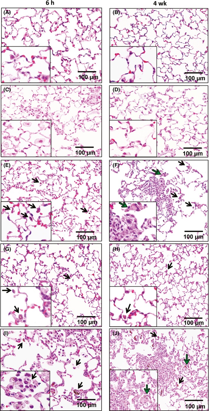
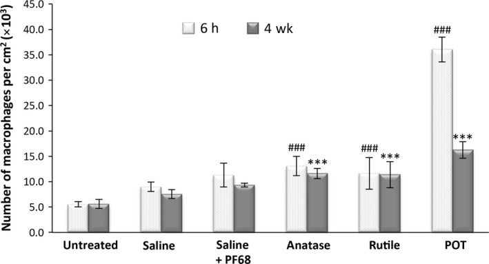
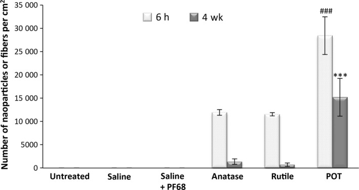
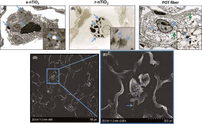
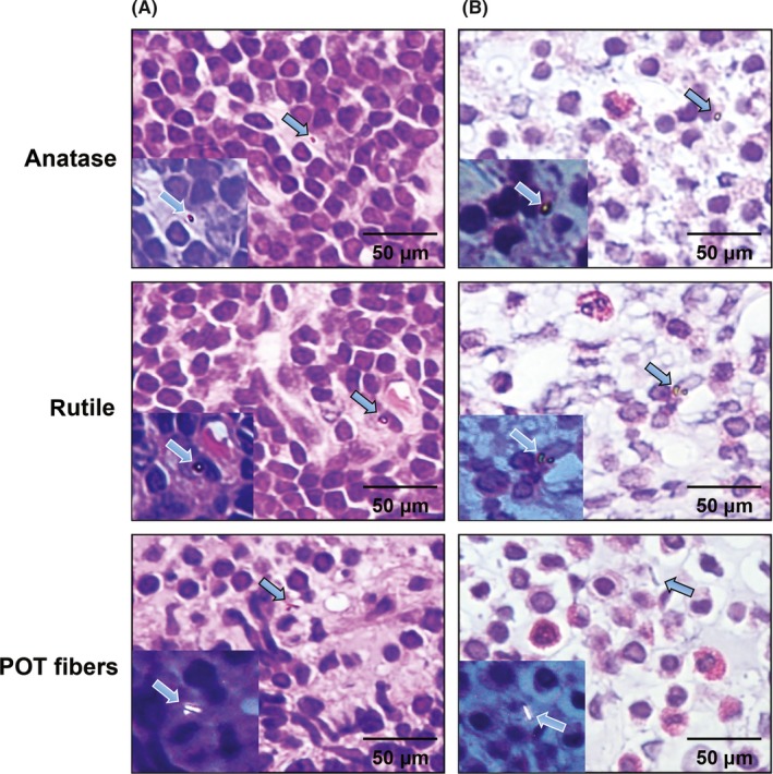
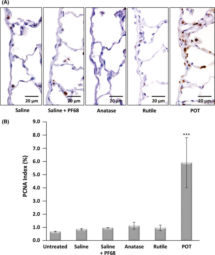
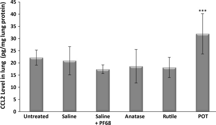
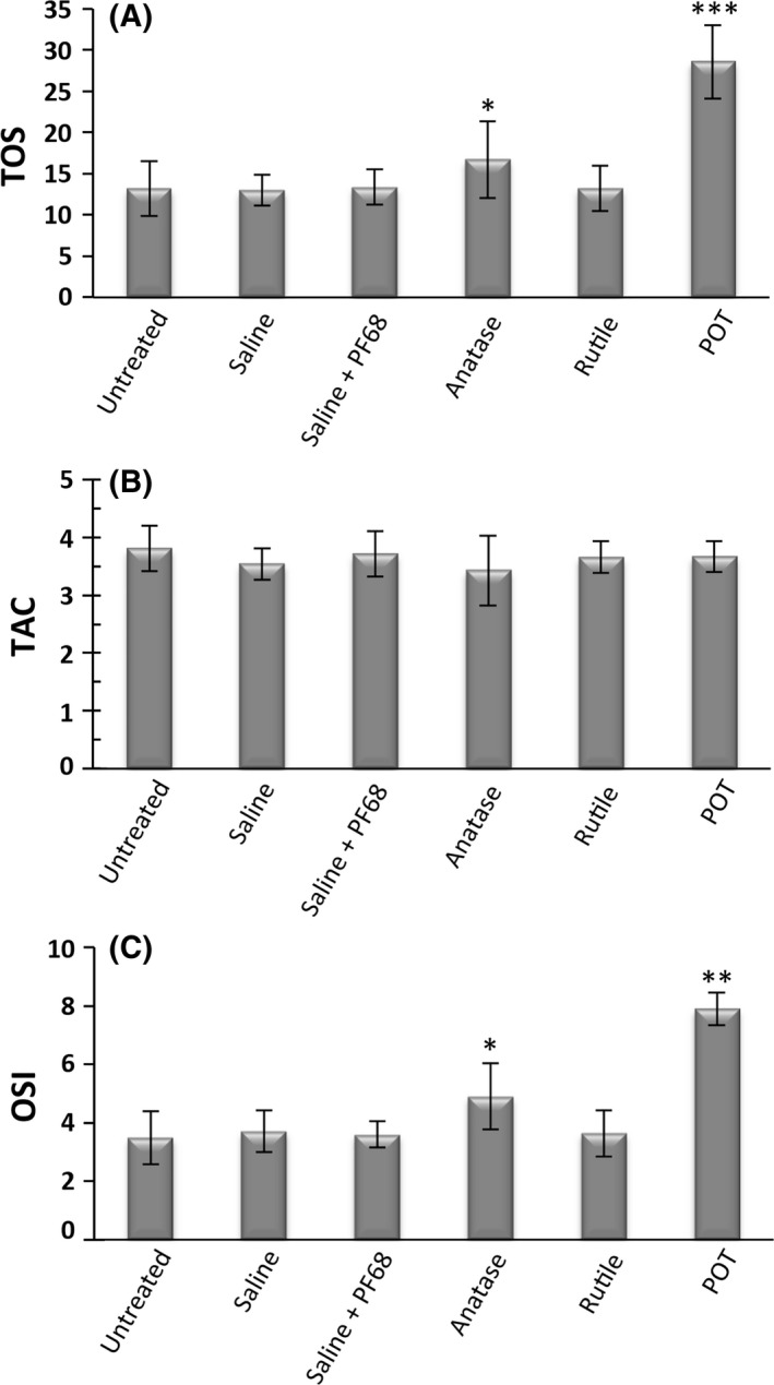
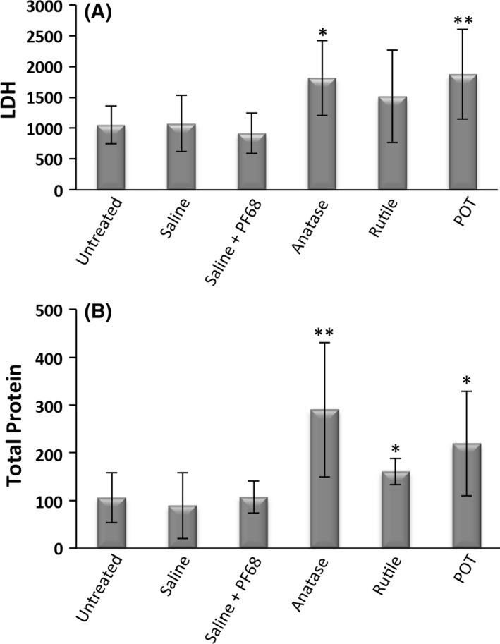
Potassium octatitanate fibers (K2 O·8TiO2 , POT fibers) are widely used as an alternative to asbestos. We investigated the pulmonary and pleural toxicity of POT fibers with reference to 2 non-fibrous titanium dioxide nanoparticles (nTiO2 ), photoreactive anatase (a-nTiO2 ) and inert rutile (r-nTiO2 ). Ten-week-old male F344 rats were given 0.5 mL of 250 μg/mL suspensions of POT fibers, a-nTiO2 , or r-nTiO2 , 8 times (1 mg/rat) over a 15-day period by trans-tracheal intrapulmonary spraying (TIPS). Rats were killed at 6 hours and at 4 weeks after the last TIPS dose. Alveolar macrophages were significantly increased in all treatment groups at 6 hours and at 4 weeks. At week 4, a-nTiO2 and r-nTiO2 were largely cleared from the lung whereas a major fraction of POT fibers were not cleared. In the bronchoalveolar lavage, alkaline phosphatase activity was elevated in all treatment groups, and lactate dehydrogenase (LDH) activity was elevated in the a-nTiO2 and POT groups. In lung tissue, oxidative stress index and proliferating cell nuclear antigen (PCNA) index were elevated in the a-nTiO2 and POT groups, and there was a significant elevation in C-C motif chemokine ligand 2 (CCL2) mRNA and protein in the POT group. In pleural cavity lavage, total protein was elevated in all 3 treatment groups, and LDH activity was elevated in the a-nTiO2 and POT groups. Importantly, the PCNA index of the visceral mesothelium was increased in the POT group. Overall, POT fibers had greater biopersistence, induced higher expression of CCL2, and provoked a stronger tissue response than a-nTiO2 or r-nTiO2 .

Keywords: inhalation toxicity; potassium octatitanate fiber; rat; titanium dioxide nanoparticle; trans-tracheal intrapulmonary spraying.


Figures

Figure 1
Figure 1
Characterization of test materials in suspension. A, Scanning electronic microscope images and B, transmission electron microscopy images of a, photoreactive anatase (a‐nTiO 2), b, inert rutile (r‐nTiO 2), and c, potassium octatitanate (POT) fibers
Figure 2
Figure 2
Histological observation of lung tissue at 6 h and at 4 wks after the final trans‐tracheal intrapulmonary spraying dose of A, B, saline, C, D, saline + PF68, E, F, photoreactive anatase (a‐nTiO 2), G, H, inert rutile (r‐nTiO 2), and I, J, potassium octatitanate (POT) fibers. Black arrows indicate alveolar macrophages phagocytizing test materials, and green arrows indicate multinucleated giant cells
Figure 3
Figure 3
Alveolar macrophage counts per cm2 of lung tissue. ### P < .001 vs vehicle (photoreactive anatase [a‐nTiO 2] and inert rutile [r‐nTiO 2] vs saline; potassium octatitanate [POT] vs saline + PF68) at 6 h, and ***P < .001 vs vehicle (a‐nTiO 2 and r‐nTiO 2 vs saline; POT vs saline + PF68) 4 wks after the final trans‐tracheal intrapulmonary spraying dose
Figure 4
Figure 4
Nanoparticle/fiber count per cm2 of lung tissue. ### P < .001 vs photoreactive anatase (a‐nTiO 2) and inert rutile (r‐nTiO 2) at 6 h and ***P < .001 vs a‐nTiO 2 and r‐nTiO 2 4 wks after the final trans‐tracheal intrapulmonary spraying dose. POT, potassium octatitanate
Figure 5
Figure 5
Transmission electron microscopy (TEM) and scanning electronic microscopy (SEM) of alveolar macrophages. (A‐C) TEM images of the 3 test materials (blue arrows) and cytoplasmic vacuoles in the macrophages phagocytosing POT fibers (green arrows). (D, E) SEM images of alveolar macrophages phagocytosing a POT fiber (arrow). a‐nTiO 2, photoreactive anatase; POT, potassium octatitanate; r‐nTiO 2, inert rutile
Figure 6
Figure 6
Translocation of the test materials. A, All 3 test materials were detected in the mediastinal lymph nodes (arrows). B, The 3 test materials were also present in the pleural cavity lavage cell pellets (arrows). POT, potassium octatitanate
Figure 7
Figure 7
Proliferating cell nuclear antigen (PCNA) staining of lung tissue. PCNA index expressed as the percentage of PCNA‐positive cells to the total number of pulmonary cells per slide. **P < .01 and ***P < .001 vs vehicle (photoreactive anatase [a‐nTiO 2] vs saline; potassium octatitanate [POT] vs saline + PF68) 4 wks after the final trans‐tracheal intrapulmonary spraying dose
Figure 8
Figure 8
Visceral mesothelial cell proliferation. A, Proliferating cell nuclear antigen (PCNA) staining of visceral mesothelial cells. B, PCNA index expressed as the percentage of PCNA‐positive cells to the total number of visceral mesothelial cells per slide. ***P < .001 vs vehicle (saline + PF68) 4 wks after the final trans‐tracheal intrapulmonary spraying dose. POT, potassium octatitanate
Figure 9
Figure 9
Proinflammatory cytokine gene expression in lung tissue. Graphs show RNA expression of the target cytokines relative to actin. *P < .05 and **P < .01 vs vehicle (inert rutile [r‐nTiO 2] vs saline; potassium octatitanate [POT] vs saline + PF68) 4 wks after the final trans‐tracheal intrapulmonary spraying dose. CCL, C‐C motif chemokine ligand
Figure 10
Figure 10
C‐C motif chemokine ligand 2 (CCL2) protein expression in lung tissue. ***P < .001 vs vehicle (saline + PF68) 4 wks after the final trans‐tracheal intrapulmonary spraying dose. POT, potassium octatitanate
Figure 11
Figure 11
Total oxidant status (TOS), total antioxidant capacity (TAC), and oxidative stress index (OSI) in lung tissue. A, TOS is expressed as μmol H2O2 equivalents per gram of tissue. B, TAC is expressed as μmol Trolox equivalents per gram of tissue. C, OSI is expressed as the ratio of TOS to TAC. *P < .05, **P < .01, and ***P < .001 vs vehicle (photoreactive anatase [a‐nTiO 2] vs saline; potassium octatitanate [POT] vs saline + PF68) 4 wks after the final trans‐tracheal intrapulmonary spraying dose
Figure 12
Figure 12
Biochemical analysis of bronchoalveolar lavage fluid. A, Lactate dehydrogenase (LDH) enzyme activity, measured as micro‐units/L and B, alkaline phosphatase (ALP) enzyme concentration, measured as units/L. *P < .05 and ***P < .001 vs vehicle (photoreactive anatase [a‐nTiO 2] and inert rutile [r‐nTiO 2] vs saline; potassium octatitanate [POT] vs saline + PF68) 4 wks after the final trans‐tracheal intrapulmonary spraying dose
Figure 13
Figure 13
Biochemical analysis of pleural cavity lavage fluid. A, Lactate dehydrogenase (LDH) enzyme activity, measured as micro‐units/L and B, total protein concentration (μg/mL). *P < .05 and **P < .01 vs vehicle (photoreactive anatase [a‐nTiO 2] and inert rutile [r‐nTiO 2] vs saline; potassium octatitanate [POT] vs saline + PF68) 4 wks after the final trans‐tracheal intrapulmonary spraying dose

Similar articles

Cited by

References

    1. Yamato H, Morimoto Y, Tsuda T, et al. Clearance of inhaled potassium Octatitanate Whisker from rat lungs. J Occup Health. 2002;44:34‐39.
    1. Donaldson K, Murphy F, Schinwald A, Duffin R, Poland CA. Identifying the pulmonary hazard of high aspect ratio nanoparticles to enable their safety‐by‐design. Nanomedicine (Lond). 2011;6:143‐156. - PubMed
    1. Adachi S, Kawamura K, Takemoto K. A trial on the quantitative risk assessment of man‐made mineral fibers by the rat intraperitoneal administration assay using the JFM standard fibrous samples. Ind Health. 2001;39:168‐174. - PubMed
    1. Stanton MF, Layard M. The carcinogenicity of fibrous minerals In: Gravatt CC, LaFleur PD, Heinrich KFJ, eds. Workshop on Asbestos: Definitions and Measurement Methods, vol. 506 Washington, DC: National Measurement Laboratory National Bureau of Standards; 1978:143‐151.
    1. Stanton MF, Layard M, Tegeris A, et al. Relation of particle dimension to carcinogenicity in amphibole asbestoses and other fibrous minerals. J Natl Cancer Inst. 1981;67:965‐975. - PubMed