. 2018 Aug;17(4):e12761.
doi: 10.1111/acel.12761. Epub 2018 May 17.

Loss of sirtuin 1 and mitofusin 2 contributes to enhanced ischemia/reperfusion injury in aged livers

Affiliations

Loss of sirtuin 1 and mitofusin 2 contributes to enhanced ischemia/reperfusion injury in aged livers

Sung Kook Chun et al. Aging Cell. 2018 Aug.

Abstract

Ischemia/reperfusion (I/R) injury is a causative factor contributing to morbidity and mortality during liver resection and transplantation. Livers from elderly patients have a poorer recovery from these surgeries, indicating reduced reparative capacity with aging. Mechanisms underlying this age-mediated hypersensitivity to I/R injury remain poorly understood. Here, we investigated how sirtuin 1 (SIRT1) and mitofusin 2 (MFN2) are affected by I/R in aged livers. Young (3 months) and old (23-26 months) male C57/BL6 mice were subjected to hepatic I/R in vivo. Primary hepatocytes isolated from each age group were also exposed to simulated in vitro I/R. Biochemical, genetic, and imaging analyses were performed to assess cell death, autophagy flux, mitophagy, and mitochondrial function. Compared to young mice, old livers showed accelerated liver injury following mild I/R. Reperfusion of old hepatocytes also showed necrosis, accompanied with defective autophagy, onset of the mitochondrial permeability transition, and mitochondrial dysfunction. Biochemical analysis indicated a near-complete loss of both SIRT1 and MFN2 after I/R in old hepatocytes, which did not occur in young cells. Overexpression of either SIRT1 or MFN2 alone in old hepatocytes failed to mitigate I/R injury, while co-overexpression of both proteins promoted autophagy and prevented mitochondrial dysfunction and cell death after reperfusion. Genetic approaches with deletion and point mutants revealed that SIRT1 deacetylated K655 and K662 residues in the C-terminus of MFN2, leading to autophagy activation. The SIRT1-MFN2 axis is pivotal during I/R recovery and may be a novel therapeutic target to reduce I/R injury in aged livers.

Keywords: aging; autophagy; hepatocytes; liver; mitochondria; mitophagy.


Figures

Figure 1
Figure 1
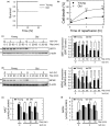
Accelerated loss of SIRT1 and MFN2 after short I/R in old livers. (a) Kaplan–Meier survival analysis of young (n = 6) and old mice (n = 10), after liver I/R in vivo. The survival probability of old mice after reperfusion was significantly lower than that of young mice (p = .025). (b) Cell death in young or old hepatocytes, after 2 hr of ischemia and 5, 60, and 120 min of reperfusion. (c–d) Immunoblotting analysis of SIRT1 and MFN2 expression after ischemia (Isch) and reperfusion (Rep) of hepatocytes. Changes in SIRT1 and MFN2 were normalized to β‐actin. (e–f) Colorimetric analysis of intracellular NAD + and NADH. I/R, ischemia/reperfusion
Figure 2
Figure 2
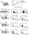
Cytoprotection by co‐overexpression of SIRT1 and MFN2. (a) Cell death after I/R of old hepatocytes. Overexpression of SIRT1 and MFN2 was induced in old hepatocytes by infecting cells with adenoviral SIRT1 (1 MOI) or MFN2 (10 MOI), or in combination. Necrotic cell death was assessed after 2 hr of ischemia. ***p < .001 vs. control. (b) Immunoblotting analysis of changes in cytosolic (C) and nuclear (N) SIRT1 during I/R. Graphs represent quantification of SIRT1 and MFN2 expression relative to subcellular markers. (c–e) Immunoblotting of SIRT1 and MFN2 expression after 2 hr of ischemia and indicated times after reperfusion
Figure 3
Figure 3
Suppression of the MPT by co‐overexpression of SIRT1 and MFN2 in old hepatocytes. (a–d) Confocal images of the onset of MPT, ΔΨm, and necrosis. Arrows indicate nuclear labeling of PI, indicative of necrotic cell death. Scale bar = 20 μm. (e) Cell death in young WT and liver‐specific SIRT1 KO hepatocytes after 2 hr of ischemia. (f) Immunoblotting analysis of MFN2 expression in young WT and SIRT1 KO hepatocytes after 1 or 2 hr of ischemia. (g) Immunoprecipitation (IP) and immunoblotting (IB) of the mitochondria‐enriched fractions from young and old hepatocytes with SIRT1 (left) or acetylated Lys (right). Graphs represent quantification of SIRT1‐MFN2 immune complexes and acetylated MFN2. (h) IP of SIRT1 and acetylated Lys in young WT and liver‐specific SIRT1 KO hepatocytes
Figure 4
Figure 4
Improved autophagy by dual overexpression of SIRT1 and MFN2. (a) Autophagic flux after 2 hr of ischemia in old hepatocytes. Autophagic flux was measured by immunoblotting LC3‐II levels in the absence or presence of 20 μm chloroquine (CQ). Graph represents quantification of LC3‐II differences before and after the addition of CQ. (b) Representative confocal images of mCherry‐GFPLC3 after I/R in young and old hepatocytes. Cells expressing control virus (LacZ) or co‐overexpressing MFN2 and SIRT1 were exposed to 2 hr of ischemia and 45 min of reperfusion. Scale bar = 10 μm. Yellow and red puncta representing autophagosomes and autolysosomes, respectively, were counted after reperfusion. (c) Autophagic flux after I/R in young WT and SIRT1 KO hepatocytes. (d) Representative confocal images of GFPLC3 and TMRM after 2 hr of ischemia followed by 30 min of reperfusion. The bottom panel is an enlarged image of the square inset. Arrows indicate the onset of mitophagy. Scale bar = 20 μm. (e) Autophagic flux in ischemic young hepatocytes after MFN2 overexpression
Figure 5
Figure 5
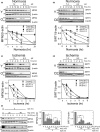
Mechanisms of MFN2 and SIRT1 depletion during I/R. (a–b) pH‐dependent changes in MFN2 and SIRT1 during normoxia in young and old hepatocytes. Immunoblotting analysis in the presence of 35 μm cycloheximide under different pH. *p < .05, **p < .01, and ***p < .001 vs. young cells at pH 7.4. # p < .05 and ## p < .01, vs. old cells at pH 7.4. (c–d) pH‐dependent changes in MFN2 and SIRT1 during anoxia in young and old hepatocytes. Effects of pH on MFN2 and SIRT1 expression were determined under anoxic condition with cycloheximide. (e) Loss of MFN2 and SIRT1 by calpains. Changes in MFN2 and SIRT1 expression were assessed with and without calpain inhibitor, ALLM (0–8 μm), after I/R in old hepatocytes. ALLM, N‐acetyl‐Leu‐Leu‐methional
Figure 6
Figure 6
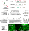
Mechanisms of SIRT1/MFN2‐mediated autophagy. (a) Schematic of MFN2 and putative SIRT1‐targeted Lys resides (K37, K215, K357, K655, K662) with cytosolic heptad repeats (HR). Diagrams of MFN2 deletion constructs. (b) Autophagic flux in MFN2 deletion mutants with SIRT1 overexpression in HEK293T cells. (c) Autophagic flux in MFN2 point mutants without SIRT1 overexpression in HEK293T cells. (d) Multiphoton images of young and old livers after I/R in vivo. After infecting with AdMFN2 and AdSIRT1, old livers were subjected to I/R in vivo. Multiphoton images of Rd‐123 were collected after 40 min of reperfusion. Scale bar = 50 μm

References

    1. Anderson, K. A. , & Hirschey, M. D. (2012). Mitochondrial protein acetylation regulates metabolism. Essays in Biochemistry, 52, 23–35. 10.1042/bse0520023 - DOI - PMC - PubMed
    1. Atkins, K. M. , Thomas, L. L. , Barroso‐González, J. , Thomas, L. , Auclair, S. , Yin, J. , … Thomas, G. (2014). The multifunctional sorting protein PACS‐2 regulates SIRT1‐mediated deacetylation of p53 to modulate p21‐dependent cell‐cycle arrest. Cell Reports, 8, 1545–1557. 10.1016/j.celrep.2014.07.049 - DOI - PMC - PubMed
    1. Bach, D. , Pich, S. , Soriano, F. X. , Vega, N. , Baumgartner, B. , Oriola, J. , … Zorzano, A. (2003). Mitofusin‐2 determines mitochondrial network architecture and mitochondrial metabolism. A novel regulatory mechanism altered in obesity. Journal of Biological Chemistry, 278, 17190–17197. 10.1074/jbc.M212754200 - DOI - PubMed
    1. Biel, T. G. , Lee, S. , Flores‐Toro, J. A. , Dean, J. W. , Go, K. L. , Lee, M. H. , … Kim, J. S. (2016). Sirtuin 1 suppresses mitochondrial dysfunction of ischemic mouse livers in a mitofusin 2‐dependent manner. Cell Death and Differentiation, 23, 279–290. 10.1038/cdd.2015.96 - DOI - PMC - PubMed
    1. de Brito, O. M. , & Scorrano, L. (2008). Mitofusin 2 tethers endoplasmic reticulum to mitochondria. Nature, 456, 605–610. 10.1038/nature07534 - DOI - PubMed

Publication types

LinkOut - more resources