. 2018 Aug;17(4):e12783.
doi: 10.1111/acel.12783. Epub 2018 May 17.

CTC1-STN1 coordinates G- and C-strand synthesis to regulate telomere length

Affiliations

CTC1-STN1 coordinates G- and C-strand synthesis to regulate telomere length

Peili Gu et al. Aging Cell. 2018 Aug.

Abstract

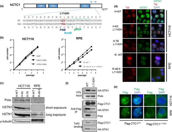
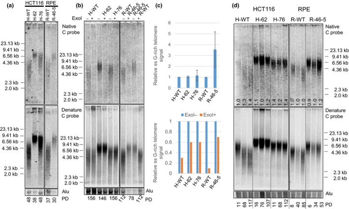
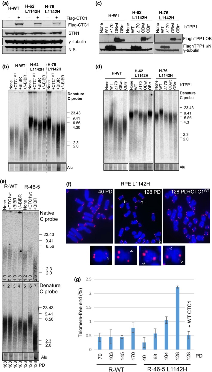
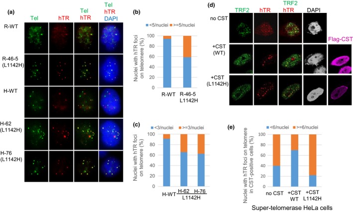
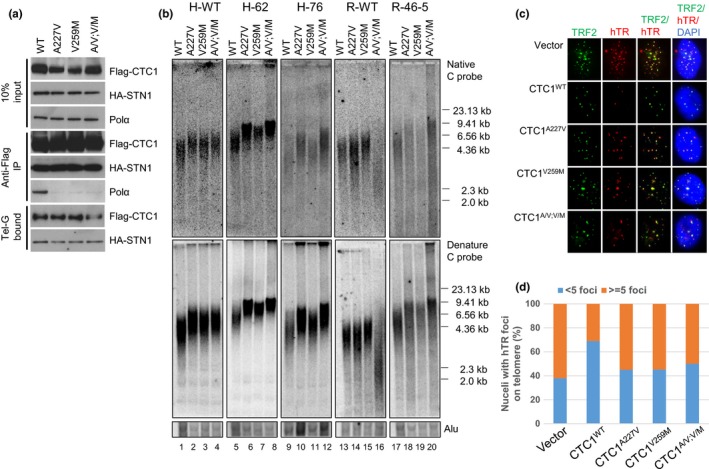
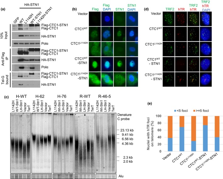
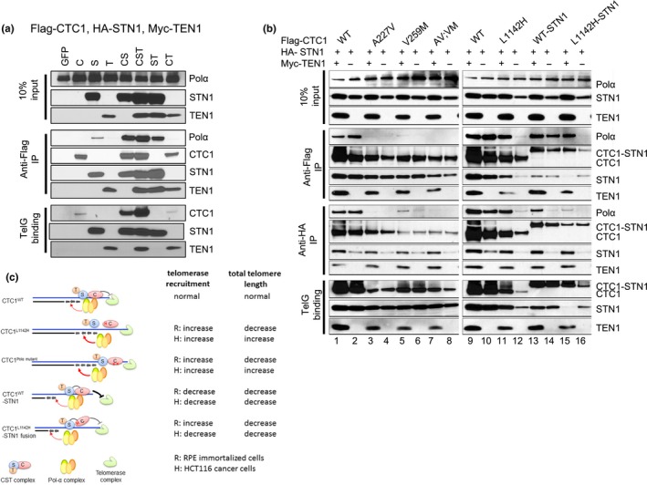
Coats plus (CP) is a rare autosomal recessive disorder caused by mutations in CTC1, a component of the CST (CTC1, STN1, and TEN1) complex important for telomere length maintenance. The molecular basis of how CP mutations impact upon telomere length remains unclear. The CP CTC1L1142H mutation has been previously shown to disrupt telomere maintenance. In this study, we used CRISPR/Cas9 to engineer this mutation into both alleles of HCT116 and RPE cells to demonstrate that CTC1:STN1 interaction is required to repress telomerase activity. CTC1L1142H interacts poorly with STN1, leading to telomerase-mediated telomere elongation. Impaired interaction between CTC1L1142H :STN1 and DNA Pol-α results in increased telomerase recruitment to telomeres and further telomere elongation, revealing that C:S binding to DNA Pol-α is required to fully repress telomerase activity. CP CTC1 mutants that fail to interact with DNA Pol-α resulted in loss of C-strand maintenance and catastrophic telomere shortening. Our findings place the CST complex as an important regulator of both G-strand extensions by telomerase and C-strand synthesis by DNA Pol-α.

Keywords: DNA repair; stem cell aging; telomerase; telomere.


Figures

Figure 1
Figure 1
Generation of the CTC1 L1142H mutation in HCT116 and RPE cells using CRISPR/Cas9. (a) Schematic of the guide sgRNA utilized to mutate CTC1L1142 to CTC1H1142. Arrows indicate PCR primers used for genotyping. (b) NIH 3T3 assays were used to measure the proliferative capacities of the indicated cell lines. (c) Expression pattern of endogenous DNA Pol‐α and STN1 in the indicated cell lines detected by western analysis. γ‐tubulin was used as a loading control. (d) Immuno‐FISH analysis for endogenous STN1 (green) and telomeres (red) in WT or L1142H mutant HCT116 or RPE cell lines. STN1 was visualized using an anti‐STN1 antibody, telomeres visualized by hybridization with a 5′‐Tam‐OO‐(CCCTAA)4‐3′ PNA probe, and nuclei visualized by 4,6‐diamidino‐2‐phenylindole staining (DAPI; blue). (e) Immunostaining for WT Flag‐CTC1 or Flag‐CTC1L1142H (green) expressed in HCT116 or RPE cells. Nuclei were stained with DAPI (in blue). (f) Co‐IP was used to determine the ability of WT Flag‐CTC1 and Flag‐CTC1L1142H mutant proteins to interact with HASTN1, endogenous DNA pol‐α, and ss telomeric DNA [Tel‐G oligo: (TTAGGG)3]
Figure 2
Figure 2
Increased telomere lengths in cells bearing the CTC1L1142H mutation. (a) TRF Southern analysis of the lengths of single‐stranded (ss) (top panel) and total telomeric DNA (bottom panel) in cells of the indicated genotypes. Numbers at the bottom indicate the number of population doublings (PDs). (b) Telomere length analysis of single‐stranded (ss) (top panel) and total telomere length (bottom panel) in cells of the indicated genotypes either treated with (+) or without (−) ExoI. Alu was used as DNA loading control. (c) Quantification of the relative ss G‐rich telomere signal normalized to total telomeric signal in cells of the indicated genotypes, either untreated (top) or treated (bottom) with Exo I. Values represent the mean from three independent experiments and error bars represent standard error of the mean (SEM). (d) Telomere length analysis of single‐stranded (ss) (top panel) and total telomere length (bottom panel) of cells of the indicated genotypes subjected to long‐term serial passaging. PD: population doublings. Alu was used as DNA loading control. Numbers in native gel refer to ratio of overhang signal intensity to total telomere intensity
Figure 3
Figure 3
CTC1 L1142H promotes telomere elongation by telomerase. (a) Determination of the expression of WT Flag‐CTC1 and endogenous STN1 in the indicated cell lines by western analysis. (b) TRF Southern analysis of telomere length in WT or mutant HCT116 cells reconstituted with either WT CTC1 or cultured in the presence of 10 μM of the telomerase inhibitor BIBR. Cells were first passaged for 120 PD, reconstituted with WT CTC1 or treated with BIBR, and maintained for another 120 PD. +/−BIBR: cells were maintained in the presence of 10 μM BIBR for 60 PD, then maintained for another 60 PD after discontinuing BIBR treatment. (C) Expression levels of WT TPP1, TPP1∆170 or TPP1‐OBWT or OBRR (K166R; K167R mutations) in HCT116 cells by western analysis. (d) Analysis of total telomeric DNA in WT or mutant HCT116 cells expressing either WT TPP1, TPP1∆170 or TPP1‐OBWT or OBRR. Cells expressed TPP1 constructs for 60 days before undergoing telomere length analysis by TRF Southern. Alu was used as a DNA loading control. (e) Telomere length analysis of single‐stranded (ss) (top panel) and total telomere length (bottom panel) in cells of the indicated genotypes. PD: population doublings. +CTC1: WT CTC1 was expressed for 120 days before the cells were harvested for telomere length analysis. +BIBR: cells were treated with 10 μM of the telomerase inhibitor BIBR. Numbers in native gel refer to ratio of overhang signal intensity to total telomere intensity. (f) Metaphase spreads revealing sister telomere loss in RPEL 1142H cells at the indicated PD. White arrowheads point to STLs. In one experiment, WT CTC1 was expressed in RPEL 1142H cells at PD 54 and then passaged for an additional 74 PD. (g) Quantification of the percentage of sister telomere loss in WT RPE or RPEL 1142H cells with the indicated PD when harvested
Figure 4
Figure 4
Increased telomerase recruitment to the telomeres of CTC1L1142H mutant cells. (a) RPE or HCT116 cells were infected with retrovirus expressing hTERT and TPP1, then transiently transfected with hTR. Fluorescence in situ hybridization (FISH) was used to detect co‐localization of hTR (red) with telomeres (5′‐Tam‐OO‐(CCCTAA)4‐3′ PNA, green) in cells of the indicated genotypes. White arrows point to co‐localized hTR‐telomere signals in nuclei. (b, c) Quantification of the percentage of hTR‐positive foci on telomeres in RPE (b) or HCT116 (c) cells. At least 100 nuclei possessing co‐localized hTR signal on telomere were counted. (d) FISH was used to detect hTR foci (red), and immunofluorescence with anti‐Flag antibody was used to detect the Flag‐CST complex (purple) and a rabbit anti‐TRF2 antibody was used to detect endogenous TRF2 (green). Telomerase recruitment to telomeres is indicated in the merge panel by yellow spots. (Magnification: 100×). (e) Quantitation of the fraction of telomerase foci‐containing cells transfected with indicated CST constructs that contained ≤5 (blue) or ≥6 (orange) hTR foci per nucleus. Number of nuclei scored: WT CTC1: 55 nuclei, mutant CTC1: 67 nuclei, telomerase alone: 66 nuclei
Figure 5
Figure 5
Disruption of CTC1:DNA Pol‐α interaction results in further telomere elongation in CTC1L1142H mutant cells. (a) Biochemical characterization of Flag‐CTC1 WT and mutants unable to interact with endogenous DNA Pol‐α. Flag‐CTC1 was incubated with HASTN1 and interaction with endogenous DNA Pol‐α was determined by Co‐IP. (b) Analysis of the lengths of the 3′ overhang (top panel) and total telomere DNA (bottom panel) in HCT116 and RPE cells expressing WT CTC1, CTC1A227V, CTC1V259M, or CTC1A227V, V259M mutants for 2 months by TRF Southern. Alu was used as a DNA loading control. (c) Expression of CTC1 mutants unable to interact with DNA Pol‐α increased telomerase recruitment to telomeres in CTC1L1142H mutant RPE cells. Cells of the indicated genotypes were infected with retrovirus expressing hTERT and TPP1, then transiently transfected with a hTR cDNA. FISH was used to detect co‐localization of hTR (red) with telomeres (anti‐TRF2 antibody, green). (d) Quantification of (c). For each cell type, a minimum of 100 nuclei with signal were scored for the number of co‐localized foci
Figure 6
Figure 6
The CTC1:STN1 complex inhibits telomerase recruitment to telomeres. (a) WT Flag‐CTC1, Flag‐CTC1L1142H, WT Flag‐CTC1 tethered to STN1 or Flag‐CTC1L1142H tethered to STN1 were examined for their ability to interact with HASTN1, endogenous DNA Pol‐α and ss Tel‐G oligo. (b) IF examination of the cellular distribution of WT Flag‐CTC1, Flag‐CTC1L1142H, WT Flag‐CTC1‐STN1, or Flag‐CTC1L1142HSTN1 in CTC1L1142H mutant RPE cells using anti‐Flag antibody (green). Blue: DAPI staining to detect nuclei. (c) TRF Southern analysis of telomere lengths in WT or CTC1L1142H mutant HCT116 or RPE cells expressing the indicated DNA constructs for 2 months. Alu was used as DNA loading control. (d) Tethering CTC1L1142H to STN1 does not inhibit telomerase recruitment to telomeres in CTC1L1142H mutant RPE cells. Cells of the indicated genotypes were infected with a retrovirus expressing hTERT and TPP1, then transiently transfected with a hTR cDNA. FISH was used to detect co‐localization of hTERC (red) with telomeres (anti‐TRF2 antibody, green). (e) Quantification of (d). A minimum of 100 nuclei for each cell type bearing hTR signals were scored for co‐localization of telomerase with telomeres
Figure 7
Figure 7
TEN1 enhances CTC1:STN1 interaction. (a) Biochemical characterization of Flag‐CTC1, HASTN1, and Myc‐TEN1 interaction with endogenous DNA Pol‐α and ss Tel‐G oligo. (b) Characterization of protein interactions between WT Flag‐CTC1, Flag‐CTC1 mutants, HASTN1, with (+) or without (−) Myc‐TEN1, with endogenous DNA Pol‐α and ss Tel‐G oligo. (c) Summary of how WT and CTC1 mutants interact with DNA Pol‐α to influence telomere binding and telomere length maintenance

References

    1. Adams, A. K. , & Holm, C. (1996). Specific DNA replication mutations affect telomere length in Saccharomyces cerevisiae . Molecular and Cellular Biology, 16, 4614–4620. 10.1128/MCB.16.9.4614 - DOI - PMC - PubMed
    1. Anderson, B. H. , Kasher, P. R. , Mayer, J. , Szynkiewicz, M. , Rice, G. I. , & Crow, Y. J. (2012). Mutations in CTC1, encoding conserved telomere maintenance component 1, cause Coats plus. Nature Genetics, 44, 338–342. 10.1038/ng.1084 - DOI - PubMed
    1. Armanios, M. , & Blackburn, E. H. (2012). The telomere syndromes. Nature Reviews Genetics, 13, 693–704. 10.1038/nrg3246 - DOI - PMC - PubMed
    1. Bhattacharjee, A. , Wang, Y. , Diao, J. , & Price, C. M. (2017). Dynamic DNA binding, junction recognition and G4 melting activity underlie the telomeric and genome‐wide roles of human CST. Nucleic Acids Research, 45, 12311–12324. 10.1093/nar/gkx878 - DOI - PMC - PubMed
    1. Bisht, K. , Smith, E. M. , Tesmer, V. M. , & Nandakumar, J. (2016). Structural and functional consequences of a disease mutation in the telomere protein TPP1. Proceedings of the National Academy of Sciences of the United States of America, 113, 13021–13026. 10.1073/pnas.1605685113 - DOI - PMC - PubMed

Publication types