. 2018 Jun 13;140(23):7046-7051.
doi: 10.1021/jacs.8b03074. Epub 2018 May 30.

Multiple Click-Selective tRNA Synthetases Expand Mammalian Cell-Specific Proteomics

Affiliations

Multiple Click-Selective tRNA Synthetases Expand Mammalian Cell-Specific Proteomics

Andrew C Yang et al. J Am Chem Soc. .

Abstract

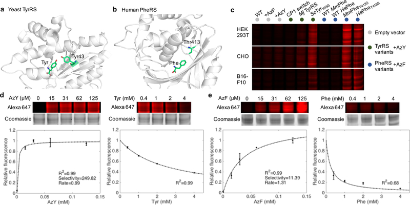
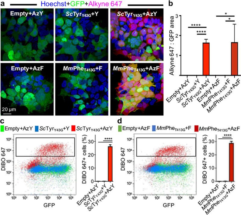
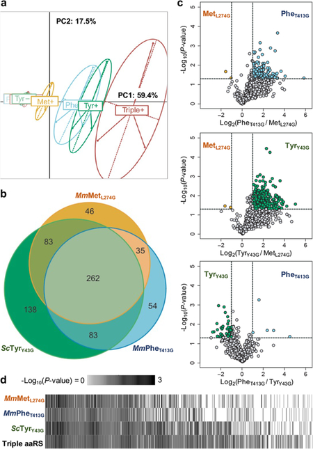
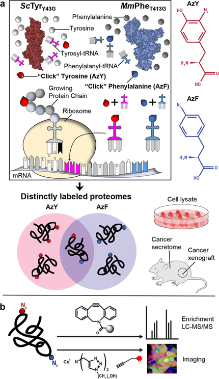
Bioorthogonal tools enable cell-type-specific proteomics, a prerequisite to understanding biological processes in multicellular organisms. Here we report two engineered aminoacyl-tRNA synthetases for mammalian bioorthogonal labeling: a tyrosyl ( ScTyrY43G) and a phenylalanyl ( MmPheT413G) tRNA synthetase that incorporate azide-bearing noncanonical amino acids specifically into the nascent proteomes of host cells. Azide-labeled proteins are chemoselectively tagged via azide-alkyne cycloadditions with fluorophores for imaging or affinity resins for mass spectrometric characterization. Both mutant synthetases label human, hamster, and mouse cell line proteins and selectively activate their azido-bearing amino acids over 10-fold above the canonical. ScTyrY43G and MmPheT413G label overlapping but distinct proteomes in human cell lines, with broader proteome coverage upon their coexpression. In mice, ScTyrY43G and MmPheT413G label the melanoma tumor proteome and plasma secretome. This work furnishes new tools for mammalian residue-specific bioorthogonal chemistry, and enables more robust and comprehensive cell-type-specific proteomics in live mammals.


Figures

Figure 1.
Figure 1.
Identification and characterization of TyrRS and PheRS variants for bioorthogonal labeling of mammalian proteomes with AzY and AzF.(a) A single substitution (Y43G) in the amino acid binding site of yeast tyrosyl-tRNA synthetase (ScTyrY43G) enables charging of the azido tyrosine analog AzY onto tRNATyr before incorporation into nascent proteins of host cells. (b) A single substitution (T413G) in the amino binding site of human or mouse phenylalanyl-tRNA synthetase (MmPheT413G) enables charging of the azido phenylalanine analog AzF onto tRNAPhe. (c) In-gel fluorescence image of Alexa Fluor 647 labeling corresponding to AzY or AzF incorporation into mammalian cell proteomes. “WT”: no exogenous amino acid added to media. “CP1 switch”: an E. coli TyrRS with the human CP1 peptide engrafted. “Mj TyRS”: TyrRS from the species M. jannaschii, without its accompanying tRNA. (d) High selectivity of ScTyrY43G for AzY over Tyr. In-gel Alexa Fluor 647 fluorescence and coomassie blue staining of whole gel lanes were quantified to assess the degree of proteome labeling normalized to total protein content. The selectivity and rate were estimated from the line of best fit to standard Michaelis−Menten kinetics at increasing AzY concentrations with 0.4 mM Tyr, and validation via increasing Tyr concentrations with 15 μM AzY. Error bars indicate standard deviation. n = 3 biological replicates. (e) As in panel d but for MmPheT413G, demonstrating high selectivity for AzF over Phe.
Figure 2.
Figure 2.
Fluorescence imaging and analysis of azide-labeled proteomes. Incorporation of the azide-bearing ncAA AzY or AzF into proteins by ScTyrY43G and MmPheT413G enables chemoselective conjugation to alkyne or DIBO-Alexa Fluor 647. Transfected HEK293T cells expressed mutant aaRS constructs or empty vector controls that coexpressed GFP, and were exposed to 125 μM AzY, AzF, tyrosine (Y), or phenylalanine (F). (a) Imaging reveals proteome labeling specific to cells expressing the mutant aaRS and exposed to AzF or AzY. Proteome labeling is pervasive across each cell. (b) Ratio of quantified Alexa Fluor 647 and GFP areas (n = 3 images). (c and d) Flow cytometry of fixed HEK293T cells (after live/dead staining), with conditions as in imaging. Only cells with mutant aaRSs and exposed to ncAAs have DIBO-AF647+ populations (n = 3 biological replicates). Error bars indicate standard deviation. *P < 0.05, ****P <0.0001.
Figure 3.
Figure 3.
Distinct proteomes labeled by each mutant aaRS. HEK293T cells transfected with equal total amounts of MmMetL274G, ScTyrY43G, and MmPheT413G, or all three aaRSs were incubated with 125 μM of their corresponding azide-bearing ncAA or endogenous amino acids. Lysates were click-enriched on DBCO beads, washed, digested, and TMT labeled before mass spectrometry. n = 3 biological replicates, in technical duplicate, for each condition. (a) Each mutant aaRS differentially labels the HEK293T proteome, as spatially represented by PCA. (b) Each mutant aaRS labels and identifies its own unique set of cell proteins. Most labeled proteins (66%, 463 of 701) are commonly detected across singly expressed mutant aaRSs, increasing confidence in their identification. (c) Among the 463 proteins identified by at least 2 aaRSs, each mutant aaRS exhibits different labeling efficiencies for different proteins. (d) The coexpression of multiple mutant aaRSs (“Triple aaRS”) enhances proteome coverage and detects more proteins with greater confidence, as assessed by P-value. Proteins significantly identified by at least one mutant aaRS were ordered by average -Log10(P-value).
Figure 4.
Figure 4.
Cell-type-specific proteome and secretome labeling in vivo. B16-F10 mouse melanoma cells stably expressing ScTyrY43G or MmPheT413G alongside GFP were implanted subcutaneously in wild-type mice, and exposed to saturating amounts of AzF, AzY, Phe, or Tyr amino acids. n = 3 mice except ScTyrY43G+Y and MmPheT413G+F, n = 2 mice. (a) In situ fluorescence confocal microscopy reveals AF647+ proteome labeling in GFP+ melanoma cells. Alkyne AF647 reacts chemoselectively to proteome-incorporated AzF and AzY. (b) Proteome-wide labeling detected via in-gel fluorescence of tumor lysates. (c) ScTyrY43G and MmPheT413G label distinct melanoma proteomes. Lysates were click-enriched on DBCO beads, washed, and digested into peptides for label-free mass spectrometry. Labeled proteomes were comprised of proteins unique to or over 5-fold more abundant than in Phe- or Tyr-exposed tumors. (d) Annotation of the labeled melanoma proteome by cellular component (STRAP), ScTyrY43G and MmPheT413G proteomes combined. (e) Ingenuity Pathway Analysis. Top pathways enriched in the melanoma proteome, ScTyrY43G and MmPheT413G combined. Multiple pathways are implicated in tumor biology. (f) ScTyrY43G and MmPheT413G label distinct melanoma plasma secretomes. (g) Pathway analysis as in panel e but for the melanoma plasma secretome.
Scheme 1.
Scheme 1.. Integrating Identifications across Mutant tRNA Synthetases Yields More Complete and Confident Proteomicsa
a(a) Engineered tRNA synthetases incorporate their azido amino acids preferentially across proteins of mammalian host cells. (b) Incorporated azide side chains are chemoselectively reacted with alkyne derivatives for protein identification and imaging.

References

    1. Conboy IM; Conboy MJ; Wagers AJ; Girma ER; Weissman IL; Rando TA Nature 2005, 433 (7027), 760–764. - PubMed
    1. Villeda SA; Plambeck KE; Middeldorp J; Castellano JM; Mosher KI; Luo J; Smith LK; Bieri G; Lin K; Berdnik D; Wabl R; Udeochu J; Wheatley EG; Zou B; Simmons DA; Xie XS; Longo FM; Wyss-Coray T Nat. Med 2014, 20 (6), 659–663. - PMC - PubMed
    1. Wyss-Coray T Nature 2016, 539, 180–186. - PMC - PubMed
    1. Nedergaard M; Verkhratsky A Glia 2012, 60 (7), 1013–1023. - PMC - PubMed
    1. Sharma K; Schmitt S; Bergner CG; Tyanova S; Kannaiyan N; Manrique-Hoyos N; Kongi K; Cantuti L; Hanisch UK; Philips MA; Rossner MJ; Mann M; Simons M Nat. Neurosci 2015, 18 (12), 1819–1831. - PMC - PubMed

Publication types

MeSH terms