. 2018 May 17;70(4):602-613.e3.
doi: 10.1016/j.molcel.2018.04.006.

C-Terminal End-Directed Protein Elimination by CRL2 Ubiquitin Ligases

Affiliations

C-Terminal End-Directed Protein Elimination by CRL2 Ubiquitin Ligases

Hsiu-Chuan Lin et al. Mol Cell. .

Abstract

The proteolysis-assisted protein quality control system guards the proteome from potentially detrimental aberrant proteins. How miscellaneous defective proteins are specifically eliminated and which molecular characteristics direct them for removal are fundamental questions. We reveal a mechanism, DesCEND (destruction via C-end degrons), by which CRL2 ubiquitin ligase uses interchangeable substrate receptors to recognize the unusual C termini of abnormal proteins (i.e., C-end degrons). C-end degrons are mostly less than ten residues in length and comprise a few indispensable residues along with some rather degenerate ones. The C-terminal end position is essential for C-end degron function. Truncated selenoproteins generated by translation errors and the USP1 N-terminal fragment from post-translational cleavage are eliminated by DesCEND. DesCEND also targets full-length proteins with naturally occurring C-end degrons. The C-end degron in DesCEND echoes the N-end degron in the N-end rule pathway, highlighting the dominance of protein "ends" as indicators for protein elimination.

Keywords: C-end degron; CRL2 ubiquitin ligase; DesCEND; protein quality control.


Conflict of interest statement

DECLRATION OF INTERESTS

The authors declare no competing interests.

Figures

Figure 1.
Figure 1.
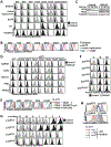
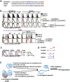
CRL2 functions in protein quality control. (A) A schematic representation of the CRL2 GPS screen. The GPS reporter system is based on the expression of two fluorescent proteins from a single promoter enabled by an internal ribosome entry site (IRES). GFP is fused to the N-terminus of the protein of interest, whereas RFP serves as an internal control. The GFP/RFP ratio represents protein stability. To search for CRL2 substrates, the GPS v5.1 HEK293T cell library was treated or not treated with DNCul2 and compared. (B) GPS reporter cells expressing putative CRL2 substrates were treated or not treated with DNCul2 and analyzed. Truncated and nonsense proteins are marked as * and NS, respectively. (C) Cycloheximide (CHX)-chase analysis of identified CRL2 substrates with or without DNCul2 treatment. (D) Western blot analysis of identified CRL2 substrates with or without DNCul2 treatment. Tubulin serves as a loading control. (E) Sequence analysis of CRL2 substrates. Protein numbers are indicated in parentheses. (F) GPS assay of truncated CRL2 substrates and their corresponding full-length versions. See also Figure S1 and Table S1.
Figure 2.
Figure 2.
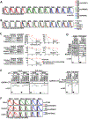
CRL2 recognizes the C-termini of aberrant proteins. (A) GPS cells expressing substrates labeled at the top with mutations indicated at left were treated or not treated with DNCul2 and analyzed. “Original” represents the original clone identified from the GPS screen. To change the protein C-terminus, the last two amino acids of substrates were deleted (A2) or the last four residues of GAPDH (ASKE) were added for capping. Due to large variations in protein stability, each GPS plot is presented with distinct ratio scaling for better resolution. As a result, the GFP/RFP ratios from different plots cannot be compared. (B) Stability comparison among proteins in (A). (C) GPS cells with or without DNCul2 virus infection were pretreated with MLN4924 for 8 hours, released, and the stability of accumulated CRL2 substrates were analyzed by CHX-chase assay. (D) Stability analysis of proteins indicated at left with or without C-terminal-tagging of the 12-residue CTT of proteins indicated at the top. (E) Stability comparisons among proteins in (D). (F) GPS assay of GAPDH with various CTTs added at its C-terminus (C), N-terminus (N) or middle (M). (G, H) Stability analysis of GAPDH C-terminally tagged with various lengths of CTTs. See also Figure S1 and Table S1.
Figure 3.
Figure 3.
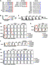
CRL2 targets aberrant proteins through various BC-box proteins. (A) GPS cells carrying indicated CRL2 substrates were treated with shRNAs against various BC-box proteins and analyzed. (B) GPS assay for cells infected with viruses expressing various BC-box proteins. (C) CHX-chase analysis of CRL2 substrates in cells with or without BC-box protein knockdown. (D) GST pull-down assay using cells expressing GST or GST-tagged BC-box proteins and GFP-tagged CRL2 substrates. (E) Physical interaction between GST-tagged BC-box proteins and GAPDH with the 12-residue CTT from various CRL2 substrates fused at its C-terminus (C), N-terminus (N) or middle (M). (F) Stability analysis of CRL2 substrates in various cells. See also Figure S2.
Figure 4.
Figure 4.
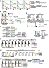
Characterization of KLHDC3, KLHDC2 and FEM1C degrons. (A) The last 12 amino acids of KLHDC3, KLHDC2 and FEM1C substrates. Critical residues demonstrated by mutagenesis are colored. The minimal lengths of some degrons were mapped and are underlined. (B, C) Stability analysis of NS19 mutants. (D-G) GPS cells expressing substrates indicated at left with mutations labeled at the top were treated with shRNAs against various BC-box proteins and analyzed. To avoid the effect of upstream Arginines, GAPDH fused with 6-amino acid CTTs from KLHDC3 substrates were tested in (E). sh#1 was used for BC-box protein knockdown unless otherwise indicated. See also Figure S3.
Figure 5.
Figure 5.
Characterization of APPBP2-mediated degradation. (A) The C-terminal sequences of APPBP2 substrates. Critical residues are colored and the minimal length of APPBP2 degrons is underlined. (B) Mutagenesis analysis of APPBP2 substrates. G-1 represents mutants that lack amino acids downstream of the critical Glycine (see Fig. S3C for stability comparisons). (C) Physical interaction between GST-tagged APPBP2 and identified (WT) or mutant APPBP2 substrates. (D) Stability analysis of KLHDC3 and APPBP2 substrate mutants with amino acids deleted or added between the critical Arginine and Glycine. Added amino acids are labeled blue. (E) Physical binding between GST-tagged WT or mutant APPBP2 with GFP-tagged APPBP2 substrates (see Fig. S3D for APPBP2 mutation). (F) GPS assay for cells expressing CRL2 substrates treated with or without shRNA against APPBP2 and infected with viruses expressing full-length (FL) or processed APP. (G) GST pull-down assay using cells expressing GST-tagged BC-box proteins and GFP-tagged CRL2 substrates with or without the presence of full-length or processed APP. See also Figure S3.
Figure 6.
Figure 6.
Physiological functions of CRL2-mediated DesCEND. (A) Competition assay between cells treated and not treated with shRNAs against various proteins involved in NMD (SMG1 and UPF1) or translation termination (RF1 and RF3) under different genetic backgrounds of BC-box proteins (see Fig. S4A for experimental procedures and Fig. S4B for results using a different shRNA). (B) Stability analysis of truncated and full-length SEPW1. (C) CHX-chase analysis of endogenous USP1 using HEK293T cells infected with viruses expressing KLHDC2, shKLHDC2 or DNCul2. Full-length (FL) and the N-terminal domain (NTD) of USP1 are labeled. (D) Physical interaction between GST-tagged BC-box proteins and GFP-tagged wild-type (WT) or mutant USP1-NTD. (E) Stability analysis of various forms of USP1 in HEK293T cells. FL-C90S is a full-length USP1 mutant that is unable to perform autocleavage. NTDAcdh1 lacks the Cdh1 degron (a.a. 295–342). (F, G) Stability analysis of GAPDH C-terminally-tagged with the CTT of USP1-NTD, ubiquitin or SUMO2. (H) Stability analysis of ubiquitin and SUMO2 with or without GS linkers. See also Figure S4.
Figure 7.
Figure 7.
DesCEND regulates full-length proteins with native C-end degrons. (A) The C-terminal sequences of indicated proteins. Critical residues in KLHDC3 degrons are colored. (B) Stability analysis of various forms of PPP1R15A, USP49 and TCAP. (C) Stability comparison among proteins in (B). (D) Characteristics of C-end degrons and their respective BC-box proteins. (E) Model of the physiological functions of DesCEND.

Comment in

References

    1. Arribere JA, Cenik ES, Jain N, Hess GT, Lee CH, Bassik MC, and Fire AZ (2016). Translation readthrough mitigation. Nature 534, 719–723. - PMC - PubMed
    1. Bachmair A, Finley D, and Varshavsky A (1986). In vivo half-life of a protein is a function of its amino-terminal residue. Science 234, 179–186. - PubMed
    1. Balch WE, Morimoto RI, Dillin A, and Kelly JW (2008). Adapting proteostasis for disease intervention. Science 319, 916–919. - PubMed
    1. Bengtson MH, and Joazeiro CA (2010). Role of a ribosome-associated E3 ubiquitin ligase in protein quality control. Nature 467, 470–473. - PMC - PubMed
    1. Bennett EJ, Rush J, Gygi SP, and Harper JW (2010). Dynamics of cullin-RING ubiquitin ligase network revealed by systematic quantitative proteomics. Cell 143, 951–965. - PMC - PubMed

Publication types