. 2018 Jul 6;430(14):2164-2180.
doi: 10.1016/j.jmb.2018.05.007. Epub 2018 May 16.

Enhanced Sampling of Interdomain Motion Using Map-Restrained Langevin Dynamics and NMR: Application to Pin1

Affiliations

Enhanced Sampling of Interdomain Motion Using Map-Restrained Langevin Dynamics and NMR: Application to Pin1

Jill J Bouchard et al. J Mol Biol. .

Abstract

Many signaling proteins consist of globular domains connected by flexible linkers that allow for substantial domain motion. Because these domains often serve as complementary functional modules, the possibility of functionally important domain motions arises. To explore this possibility, we require knowledge of the ensemble of protein conformations sampled by interdomain motion. Measurements of NMR residual dipolar couplings (RDCs) of backbone HN bonds offer a per-residue characterization of interdomain dynamics, as the couplings are sensitive to domain orientation. A challenge in reaching this potential is the need to interpret the RDCs as averages over dynamic ensembles of domain conformations. Here, we address this challenge by introducing an efficient protocol for generating conformational ensembles appropriate for flexible, multi-domain proteins. The protocol uses map-restrained self-guided Langevin dynamics simulations to promote collective, interdomain motion while restraining the internal domain motion to near rigidity. Critically, the simulations retain an all-atom description for facile inclusion of site-specific NMR RDC restraints. The result is the rapid generation of conformational ensembles consistent with the RDC data. We illustrate this protocol on human Pin1, a two-domain peptidyl-prolyl isomerase relevant for cancer and Alzheimer's disease. The results include the ensemble of domain orientations sampled by Pin1, as well as those of a dysfunctional variant, I28A-Pin1. The differences between the ensembles corroborate our previous spin relaxation results that showed weakened interdomain contact in the I28A variant relative to wild type. Our protocol extends our abilities to explore the functional significance of protein domain motions.

Keywords: domain motion; ensemble description; multi-conformational fitting; residual dipolar couplings; self-guided Langevin dynamics.


Figures

Fig. 1
Fig. 1
Human Pin1, a two-domain mitotic regulator with PPIase (cyan) and WW (magenta) domains; the structure is a snapshot from a 30ns MapSGLD-NMR simulation started from a conformer of 1NMV . The –Z and –X axes are the principal axes of PPIase domain inertia tensor (origin at its center of mass). Linker flexibility (green curved arrow) enables relative domain motion. Residue I28 lies in the WW domain (Yellow), the site of the I28A substitution. The strategy for simulating interdomain motion is: (i) strong Cartesian restraints hold the PPIase fixed in space, (ii) while the WW domain moves as a nearly rigid body by map restraints (green curved arrow).
Fig. 2
Fig. 2
(A) RDC Q factors (blue and maroon bars) for assessing agreement between 1H-15N RDCs predicted from AMBER generated conformations with those from the NMR experiments. The Q factors are averages are over the three parallel production-run simulations started from structures 1NMV-1, 1NMV-2, and 1NMV-4 from the solution structure deposition PDB 1NMV ; the error bars are the corresponding standard deviations of Q factors. The predicted RDCs came from singular value decomposition (SVD) fitting to the experimental RDCs , using the “ –bestFit ” option in PALES . Pairs of blue and maroon bars indicate Q factors for 4% C8E5/octanol media (blue), and 10 mg/ml Pf1 phage (maroon), respectively. Q factors for full-length Pin1, the WW domain, and the PPIase domain are indicated. The dashed-dotted line distinguishes two sets of Q factors. Left of the line are Q-factors just after energy minimization and before the production-run simulations. Right of the line are the analogous Q factors after 10 ns of the three production-run AMBER simulations, using the MapSGLD-NMR protocol. The reduction in Q factors after 10 ns (1/3 of the total simulation time per starting structure) is apparent. (B, C) Representative correlation plots between the 1H-15N RDCs predicted by AMBER conformations (x-axes) versus experiment (y-axes). Black circles are RDCs in and 4% C8E5; red squares are RDCs in 10 mg/ml Pf1 phage. In (B), the standard linear regression coefficient is r = 0.88 ± 0.07 for 4% C8E5 (black circles) and r = 0.87 ± 0.03 for 10 mg/ml Pf1 phage (red squares); (C) after 10 nanoseconds of production-run simulations, the standard linear regression coefficient is r = 0.991 ± 0.002 for 4% C8E5 (black circles) and r = 0.984 ± 0.008 for 10 mg/ml Pf1 phage (red squares).
Fig. 3
Fig. 3
Identification of reduced conformational ensembles using the genetic algorithm (GA) approach for multi-conformational fit . The bars indicate conformations with a fractional population > 0.001 for WT-Pin1 (A) and the single-substitution mutant, I28A-Pin1 (C). Linear correlation plots of predicted versus experimental 1H-15N RDCs recorded in 4% C8E5 media for WT-Pin1 (B) and I28A-Pin1 (D). Open circles (○) are RDCs predicted by the multi-conformational fit (WT QM = 0.196, I28A-Pin1 QM = 0.202); turquoise crosses ✚ are RDCs predicted by the GA ensemble conformation yielding the lowest single conformer Q factor (best single conformational fit): WT-Pin1 Q = 0.236, and I28A-Pin1 Q = 0.280.
Fig. 4
Fig. 4
WT-Pin1 conformations sampled by three 30ns AMBER simulations, using the MapSGLD-NMR protocol. The simulations started from three different structures (1NMV-1, 1NMV-2, and 1NMV-3) producing a total of 90,000 conformations. (A) The relative domain orientation is specified by an interdomain vector (green arrow) that starts at the geometric center of the PPIase domain active site and ends at the geometric center of the WW domain substrate-binding loop (residues 16-21). The interdomain vector coordinates (length r and angles θ and ϕ) are in the principle axis frame of the PPIase domain inertia tensor. (B) The six conformers identified by the GA/Multi-conformational search, and their corresponding interdomain vectors (green arrows defined in (A)). Panels (C–E) show histograms for the spherical coordinates of the interdomain vector, including (C) the radial (distance) coordinate r; (D) the polar angle θ; and (E) the azimuthal angle ϕ. Panel (F) is the average and the radius of gyration. Black bars represent the 90,000 conformations of the raw WT MapSGLD-NMR ensemble; the red trace represents the analogous 90,000 conformations for the raw I28A MapSGLD-NMR ensemble. Green diamonds (◆) indicate the six WT conformers; red x’s ( formula image
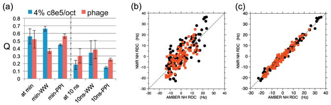
) indicate the seven I28A conformers identified by GA/multi-conformational search; stacked diamonds/x’s are populations in nearly the same bins. Panels (G and H) compare conformations sampled by the raw WT-Pin1 versus I28A-Pin1 ensembles (i.e. MapSGLD-NMR simulations producing 90,000 conformations). Yellow-orange ribbons indicate the most frequently sampled conformation; magenta indicates a less populated conformation.
Fig. 5
Fig. 5
2-d histograms comparing the conformational space sampled by (A) 20 nanoseconds rigid-body Map-SGLD-NMR simulations, implicit solvent model, and map and RDC restraints enforced; (B) 20 nanoseconds Map-SGLD simulation, implicit solvent model, map restraints enforced but RDC restraints absent; (C) depicts 400 nanoseconds explicit-solvent simulations lacking both map and RDC restraints. Comparison of (A) and (B) indicates certain conformations are excluded by the RDC data; Comparison of (B) and (C) indicates many conformations are not sampled in the explicit-solvent simulation. All simulations were started from the same structure, marked by the star.
Fig. 6
Fig. 6
Comparison of interdomain vector orientations ( r, Θ, ϕ) sampled by WT-Pin1 and I28A-Pin1. The PPIase domains are superimposed and fixed (cyan); the black lines are the –X and –Z principal axes of its inertia tensor. The dots represent the tips of the interdomain vectors starting at the geometric center of the PPIase domain active site, and terminating at the geometric center of the WW domain-binding loop. Blue dots are the WT-Pin1 ensemble; gold dots are the I28A-Pin1 ensemble. The interdomain vectors (red lines) and WW domains (dark blue) for conformers 4 and 6 from the WT GA ensemble are shown.

References

    1. Pufall MA, Graves BJ. Autoinhibitory domains: modular effectors of cellular regulation. Annu Rev Cell Dev Biol. 2002;18:421–62. - PubMed
    1. Bhattacharyya RP, Remenyi A, Yeh BJ, Lim WA. Domains, motifs, and scaffolds: The role of modular interactions in the evolution and wiring of cell signaling circuits. Annual Review of Biochemistry. 2006:655–80. - PubMed
    1. Lim WA. Designing customized cell signalling circuits. Nature Reviews Molecular Cell Biology. 2010;11:393–403. - PMC - PubMed
    1. Bax A. Weak alignment offers new NMR opportunities to study protein structure and dynamics. Protein Sci. 2003;12:1–16. - PMC - PubMed
    1. Tolman JR, Flanagan JM, Kennedy MA, Prestegard JH. NMR evidence for slow collective motions in cyanometmyoglobin. Nature Structural Biology. 1997;4:292–7. - PubMed

Publication types

Substances

LinkOut - more resources