

A study of the factors associated with cervical spinal disc degeneration, with a focus on bone metabolism and amino acids, in the Japanese population: a cross sectional study
- PMID: 29776411
- PMCID: PMC5960180
- DOI: 10.1186/s12891-018-2055-1
A study of the factors associated with cervical spinal disc degeneration, with a focus on bone metabolism and amino acids, in the Japanese population: a cross sectional study
Abstract
Background: The physical and biochemical factors responsible for cervical disc degeneration, and resulting in various spinal disorders, remain unclear. This study aimed to evaluate the correlation between cervical spinal canal stenosis and degeneration of intervertebral discs, and to analyze the factors related to disc degeneration in the Japanese population.
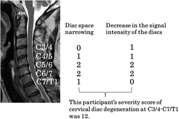
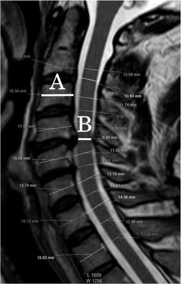
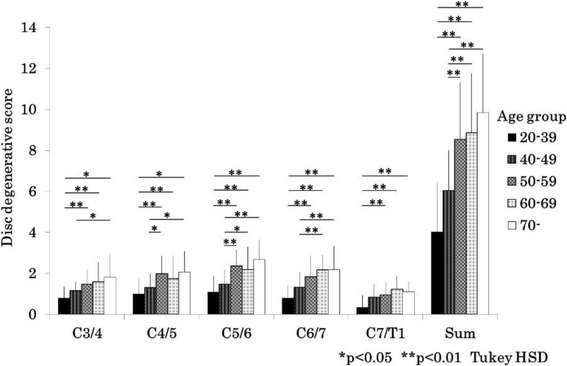
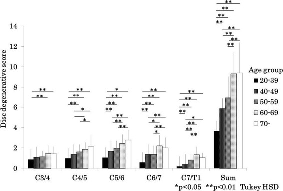
Methods: Three hundred and forty-four Japanese general residents underwent investigations, including magnetic resonance imaging of the cervical spine, in our health check project. We measured anteroposterior diameters at the levels of the cervical spinal disc in mid sagittal plane magnetic resonance imaging and evaluated disc degeneration. Spearman correlation coefficient was used to evaluate whether the diameters were correlated with disc degenerative scores. Stepwise multiple linear regression analysis was conducted with the score of disc degeneration as the dependent variable; and age, physical measurement values, bone mineral density of the forearm, and the value of serum bone metabolic markers and amino acids as the independent variables for each sex.
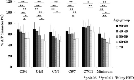
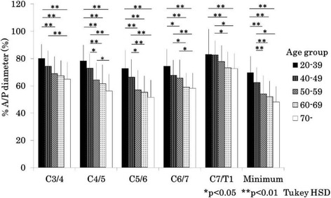
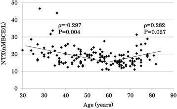
Results: As the age increased, the anteroposterior diameters decreased in both sexes. The minimum anteroposterior diameters were correlated with the disc degenerative scores (Spearman r = - 0.59, p < 0.001 in men, Spearman r = - 0.53, p < 0.001 in women). In multiple linear regression analysis, age, cross-linked N-telopeptide of type 1 collagen and isoleucine were significantly correlated with the cervical disc degenerative score in men (R2 = 0.47), and age and lysine were significantly correlated with the degenerative score in women (R2 = 0.50).
Conclusion: The factors responsible for cervical disc degeneration differed between men and women. Whether modifying these significant factors is possible, or whether this intervention would contribute to prevention of disc degeneration requires future studies.
Keywords: Amino acid; Bone metabolism; Cervical spine; Disc degeneration.
Conflict of interest statement
Ethics approval and consent to participate
For this cross-sectional survey, the ethics committee of Hirosaki University, Graduate School of Medicine approved the study (2014–377), and all participants provided written informed consent before participation.
Competing interests
The authors declare that they have no competing interests.
Publisher’s Note
Springer Nature remains neutral with regard to jurisdictional claims in published maps and institutional affiliations.
Figures
Similar articles
-
Associations between cervical disc degeneration and muscle strength in a cross-sectional population-based study.PLoS One. 2019 Jan 25;14(1):e0210802. doi: 10.1371/journal.pone.0210802. eCollection 2019. PLoS One. 2019. PMID: 30682082 Free PMC article.
-
Normative Magnetic Resonance Imaging Data of Age-Related Degenerative Changes in Cervical Disc Morphology.World Neurosurg. 2021 Aug;152:e502-e511. doi: 10.1016/j.wneu.2021.05.123. Epub 2021 Jun 16. World Neurosurg. 2021. PMID: 34098133
-
Age-related changes of thoracic and cervical intervertebral discs in asymptomatic subjects.Spine (Phila Pa 1976). 2010 Jun 15;35(14):1359-64. doi: 10.1097/BRS.0b013e3181c17067. Spine (Phila Pa 1976). 2010. PMID: 20505574
-
Magnetic resonance imaging assessment of degenerative cervical myelopathy: a review of structural changes and measurement techniques.Neurosurg Focus. 2016 Jun;40(6):E5. doi: 10.3171/2016.3.FOCUS1667. Neurosurg Focus. 2016. PMID: 27246488 Review.
-
Association between Modic changes, disc degeneration, and neck pain in the cervical spine: a systematic review of literature.Spine J. 2020 May;20(5):754-764. doi: 10.1016/j.spinee.2019.11.002. Epub 2019 Nov 13. Spine J. 2020. PMID: 31731008
Cited by
-
Biomechanical Analysis of the Reasonable Cervical Range of Motion to Prevent Non-Fusion Segmental Degeneration After Single-Level ACDF.Front Bioeng Biotechnol. 2022 Jun 16;10:918032. doi: 10.3389/fbioe.2022.918032. eCollection 2022. Front Bioeng Biotechnol. 2022. PMID: 35782514 Free PMC article.
-
Associations between cervical disc degeneration and muscle strength in a cross-sectional population-based study.PLoS One. 2019 Jan 25;14(1):e0210802. doi: 10.1371/journal.pone.0210802. eCollection 2019. PLoS One. 2019. PMID: 30682082 Free PMC article.
-
Bone mineral density, cervical spine degeneration, head and neck posture, and neck pain in the post-menopausal females: A pilot study.PLoS One. 2021 Sep 20;16(9):e0257735. doi: 10.1371/journal.pone.0257735. eCollection 2021. PLoS One. 2021. PMID: 34543361 Free PMC article.
-
Radiographic cervical spine degenerative findings: a study on a large population from age 18 to 97 years.Eur Spine J. 2021 Feb;30(2):431-443. doi: 10.1007/s00586-020-06615-0. Epub 2020 Oct 6. Eur Spine J. 2021. PMID: 33025192
-
Causal Relationship Between Cerebrospinal Fluid Metabolites and Intervertebral Disc Disease: A Bidirectional Mendelian Randomization Study.Diagnostics (Basel). 2025 Jun 16;15(12):1526. doi: 10.3390/diagnostics15121526. Diagnostics (Basel). 2025. PMID: 40564845 Free PMC article.
References
-
- Iwasaki M, Kawaguchi Y, Kimura T, Yonenobu K. Long-term results of expansive laminoplasty for ossification of the posterior longitudinal ligament of the cervical spine: more than 10 years follow up. J Neurosurg. 2002;96:180–189. - PubMed
MeSH terms
Substances
Grants and funding
LinkOut - more resources
Full Text Sources
Other Literature Sources
Medical
Research Materials
