. 2018 May 18;15(1):150.
doi: 10.1186/s12974-018-1193-6.

Valproic acid attenuates traumatic spinal cord injury-induced inflammation via STAT1 and NF-κB pathway dependent of HDAC3

Affiliations

Valproic acid attenuates traumatic spinal cord injury-induced inflammation via STAT1 and NF-κB pathway dependent of HDAC3

Shoubo Chen et al. J Neuroinflammation. .

Abstract

Background: Microglial polarization with M1/M2 phenotype shifts and the subsequent neuroinflammatory responses are vital contributing factors for spinal cord injury (SCI)-induced secondary injury. Nuclear factor-κB (NF-κB) is considered the central transcription factor of inflammatory mediators, which plays a crucial role in microglial activation. Lysine acetylation of STAT1 seems necessary for NF-kB pathway activity, as it is regulated by histone deacetylases (HDACs). There have been no studies that have explained if HDAC inhibition by valproic acid (VPA) affects the NF-κB pathway via acetylation of STAT1 dependent of HDAC activity in the microglia-mediated central inflammation following SCI. We investigated the potential molecular mechanisms that focus on the phenotypic transition of microglia and the STAT1-mediated NF-κB acetylation after a VPA treatment.

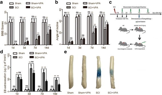
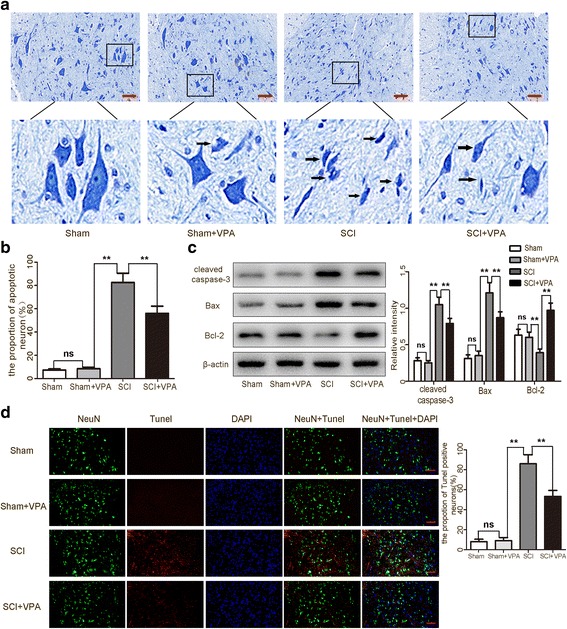
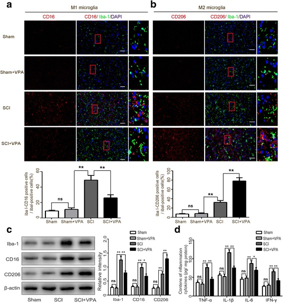
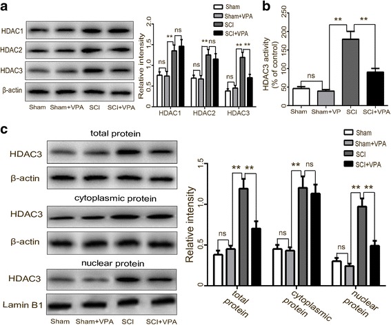
Methods: The Basso-Beattie-Bresnahan locomotion scale, the inclined plane test, the blood-spinal cord barrier, and Nissl staining were employed to determine the neuroprotective effects of VPA treatment after SCI. Assessment of microglia polarization and pro-inflammatory markers, such as tumor necrosis factor (TNF)-α, interleukin (IL)-1β, IL-6, and interferon (INF)-γ was used to evaluate the neuroinflammatory responses and the anti-inflammatory effects of VPA treatment. Immunofluorescent staining and Western blot analysis were used to detect HDAC3 nuclear translocation, activity, and NF-κB signaling pathway activation to evaluate the effects of VPA treatment. The impact of STAT1 acetylation on NF-kB pathway and the interaction between STAT1 and NF-kB were assessed to evaluate anti-inflammation effects of VPA treatment and also whether these effects were dependent on a STAT1/NF-κB pathway to gain further insight into the mechanisms underlying the development of the neuroinflammatory response after SCI.

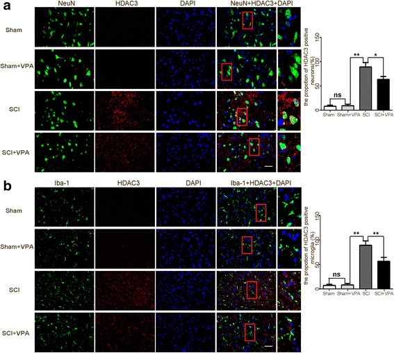
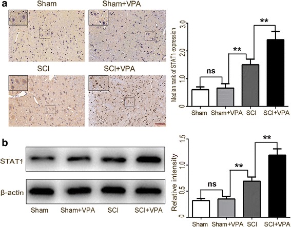
Results: The results showed that the VPA treatment promoted the phenotypic shift of microglia from M1 to M2 phenotype and inhibited microglial activation, thus reducing the SCI-induced inflammatory factors. The VPA treatment upregulation of the acetylation of STAT1/NF-κB pathway was likely caused by the HDAC3 translocation to the nucleus and activity. These results indicated that the treatment with the VPA suppressed the expression and the activity of HDAC3 and enhanced STAT1, as well as NF-κB p65 acetylation following a SCI. The acetylation status of NF-kB p65 and the complex with NF-κB p65 and STAT1 inhibited the NF-kB p65 transcriptional activity and attenuated the microglia-mediated central inflammatory response following SCI.

Conclusions: These results suggested that the VPA treatment attenuated the inflammatory response by modulating microglia polarization through STAT1-mediated acetylation of the NF-κB pathway, dependent of HDAC3 activity. These effects led to neuroprotective effects following SCI.

Keywords: HDAC3; Inflammatory; Microglia; NF-κB pathway; STAT1; Spinal cord injury; Valproic acid.


Conflict of interest statement

Ethics approval

The experimental protocols in the present study including all the surgical procedures and animal usages conformed to the guidelines for the care and use of laboratory animals by the National Institutes of Health (NIH) and were approved by the Fujian Medical University Experimental Animal Ethics Committee (Fuzhou, China).

Competing interests

The authors declare that they have no competing interests.

Publisher’s Note

Springer Nature remains neutral with regard to jurisdictional claims in published maps and institutional affiliations.

Figures

Fig. 1
Fig. 1
Neuroprotective effects of valproic acid on SCI. a Animals in the sham and sham + VPA groups obtained similar scores at the corresponding time points (score = 21). The neurological functions were severely impaired immediately after the SCI (P < 0.05). The BBB scores of these animals gradually returned to the control values. The most significant improvements were observed in the rats in the SCI + VPA group, 7 days after the SCI (8.17 ± 0.45 vs 5.03 ± 0.39) (P < 0.05). b The maximum angles were higher in SCI + VPA group than in the SCI group (27.44 ± 2.48 vs 19.75 ± 1.62) (P < 0.05). c Experimental scheme of VPA treatment after SCI. VPA was administered via intraperitoneal injection immediately following the SCI at level T10. d, e The SCI group obtained more Evans blue dye extravasation from 7 days after the SCI than the sham and the sham + VPA groups (P < 0.05). The SCI + VPA group had significantly less extravasation of Evans blue dye (P < 0.05) than the SCI group. Representative photos of the Evans blue dye extravasation in the experimental groups 7 days after the SCI. Values were expressed as mean ± standard deviation (n = 6 per group). N.S., P > 0.05, *P < 0.05, **P < 0.01
Fig. 2
Fig. 2
VPA protects neurons against SCI-induced neuronal apoptosis in the lesioned spinal cord 7 days after SCI. a, b The sham group and the sham + VPA group obtained a low apoptotic fraction of neurons 7 days after the SCI. The percentage of apoptotic cells was higher in the SCI than in the sham group (P < 0.05). The apoptotic fraction was significantly lower in the SCI + VPA than in the SCI group (P < 0.05). Representative photomicrographs of the Nissl-stained neurons are shown. Enlarged images of boxed areas are shown below. The arrows indicate the apoptotic neurons. c Western blot analyses revealed that the SCI resulted in the upregulation of apoptotic factors in the lesioned spinal cord 7 days after the SCI. The cleaved caspase-3 and Bax levels decreased and the anti-apoptotic factor Bcl-2 increased in the SCI + VPA group more than the SCI group (P < 0.05). d The TUNEL staining demonstrated that the TUNEL-positive neurons decreased significantly more in the VPA + SCI group than in the SCI group. Representative photomicrographs of the TUNEL-positive neurons are shown. The arrows indicate the apoptotic neurons. Values were expressed as mean ± standard deviation (n = 6 per group). N.S., P > 0.05, *P < 0.05, **P < 0.01. Scale bars = 50 μm
Fig. 3
Fig. 3
VPA promotes microglia polarization toward M2 and alleviates microglia-mediated inflammatory response. a, b The double immunohistochemical staining for the microglia (Iba1+) and the M1-associated marker (CD16+) or the M2-associated marker (CD206+) 7 days after the SCI was assessed. The microglia labeled with the M1 (CD16+) increased after the SCI but significantly decreased following the VPA treatment. The M2 (CD206+) increased following the VPA treatment. Representative photomicrographs of the CD16 or the CD206-positive microglia are shown. Enlarged images of boxed areas are shown on the right. c Western blot analysis showed that the M1 phenotype microglial proteins (CD16 and Iba-1) were significantly inhibited, whereas the M2 phenotype microglial protein (CD206) increased following the VPA treatment. d The VPA treatment significantly decreased the SCI-induced enhancements of TNF-α, IL-1β, IL-6, and IFN-γ (P < 0.05). Values were expressed as mean ± standard deviation (n = 6 per group). N.S., P > 0.05, *P < 0.05, **P < 0.01. Scale bars = 50 μm
Fig. 4
Fig. 4
VPA inhibits HDAC3 expression in the lesioned spinal cord. a Western blot analysis demonstrated that the expressions of HDAC 1–3 were expressed at a low level in the sham group, but were upregulated in the lesioned spinal cord 7 days after the SCI. The VPA treatment reduced the HDAC3 protein expression more than the SCI group (P < 0.05), without affecting the HDAC1 and the HDAC2 protein expressions (P > 0.05). b The HDAC3 activity in the SCI + VPA group was significantly less than in the SCI group (P < 0.05). c The expressions of HDAC3 in the cytosol, nuclei, and in total protein levels of the cells from the lesioned cortices increased after the SCI. The VPA treatment effectively decreased the HDAC3 expression in the nuclear and in the total protein of cells from the lesioned spinal cord (P < 0.05) but not in the cytosol protein (P > 0.05). Values were expressed as mean ± standard deviation (n = 6 per group). N.S., P > 0.05, *P < 0.05, **P < 0.01
Fig. 5
Fig. 5
HDAC3 expression was inhibited in both neurons and microglia after VPA treatment. Double immunohistochemical staining was used to assess the neuron (NeuN+) and the microglia (Iba-1+) in the lesioned spinal cord 7 days after the SCI. a The SCI enhanced the expression of the HDAC3 in neurons (NeuN+), which significantly decreased after the VPA treatment. Representative photomicrographs of the HDAC3-positive neurons are shown. Enlarged images of boxed areas are shown on the right. b The SCI enhanced the expression of HDAC3 in microglia (Iba-1+), which was significantly decreased following the VPA treatment. Representative photomicrographs of HDAC3-positive microglia are shown. Enlarged images of boxed areas are shown on the right. Scale bars = 50 μm
Fig. 6
Fig. 6
VPA treatment elevates STAT1 expression in the lesioned spinal cord 7 days after SCI. a STAT1 expressions were higher in the SCI than in the sham group. The STAT1 protein levels were upregulated following the VPA treatment (P < 0.05). Enlarged images of boxed areas are shown on the upper left. b Western blot analysis showed that the STAT1 protein levels were upregulated after the VPA treatment (P < 0.05). Values were expressed as mean ± standard deviation (n = 6 per group). N.S., P > 0.05, *P < 0.05, **P < 0.01. Scale bars = 50 μm
Fig. 7
Fig. 7
VPA treatment suppressed the NF-κB pathway via elevating STAT1 expression in the lesioned spinal cord 7 days after SCI. a Co-IP analysis showed a greater elevation in the STAT1 after the VPA treatment than in the SCI group (P < 0.05). b Co-IP analysis showed a greater elevation in the NF-κB acetylation after the VPA treatment than in the SCI group (P < 0.05). c The acetylated STAT1 formed a complex with the nuclear NF-κB p65. The VPA induced significant interactions between STAT1 and NF-κB p65 after the SCI (P < 0.05). d Significantly, less NF-κB p65 DNA-binding activity was observed in the SCI + VPA group than in the SCI group. e The VPA treatment inhibited NF-κB p65 nuclear translocation and expression (P < 0.05). f The inhibitory effect of the VPA treatment on the neuroinflammatory response was reversed by the pharmacological inhibition of STAT1 (fludarabine, flu). g The inhibitory effect of the VPA treatment on the NF-κB pathway was reversed by pharmacological inhibition of STAT1. Values were expressed as mean ± standard deviation (n = 6 per group). N.S., P > 0.05, *P < 0.05, **P < 0.01

References

    1. Li HT, Zhao XZ, Zhang XR, Li G, Jia, et al. Exendin-4 enhances motor function recovery via promotion of autophagy and inhibition of neuronal apoptosis after spinal cord injury in rats. Mol Neurobiol. 2016;53(6):4073–4082. doi: 10.1007/s12035-015-9327-7. - DOI - PubMed
    1. He Z, Zou S, Yin J, Gao Z, Liu Y, et al. Inhibition of endoplasmic reticulum stress preserves the integrity of blood-spinal cord barrier in diabetic rats subjected to spinal cord injury. Sci Rep. 2017;7(1):7661. doi: 10.1038/s41598-017-08052-4. - DOI - PMC - PubMed
    1. Jiang X, Yu M, Ou Y, Cao Y, Yao Y, et al. Downregulation of USP4 promotes activation of microglia and subsequent neuronal inflammation in rat spinal cord after injury. Neurochem Res. 2017;42(11):3245–3253. doi: 10.1007/s11064-017-2361-2. - DOI - PubMed
    1. Zhang Q, Bian G, Chen P, Liu L, Yu C, et al. Aldose reductase regulates microglia/macrophages polarization through the cAMP response element-binding protein after spinal cord injury in mice. Mol Neurobiol. 2016;53(1):662–676. doi: 10.1007/s12035-014-9035-8. - DOI - PubMed
    1. Fang P, Pan HC, Lin SL, Zhang WQ, Rauvala H, et al. HMGB1 contributes to regeneration after spinal cord injury in adult zebrafish. Mol Neurobiol. 2014;49(1):472–483. doi: 10.1007/s12035-013-8533-4. - DOI - PubMed

MeSH terms