

In Vivo Characterization and Quantification of Neurofibrillary Tau PET Radioligand 18F-MK-6240 in Humans from Alzheimer Disease Dementia to Young Controls
- PMID: 29777006
- PMCID: PMC6354223
- DOI: 10.2967/jnumed.118.209650
In Vivo Characterization and Quantification of Neurofibrillary Tau PET Radioligand 18F-MK-6240 in Humans from Alzheimer Disease Dementia to Young Controls
Abstract
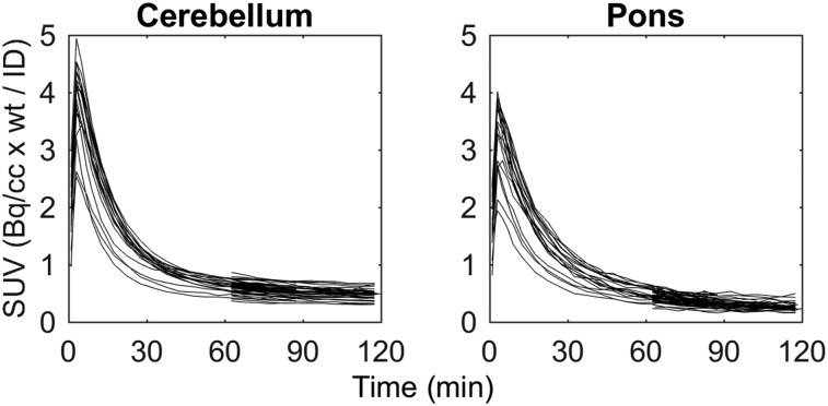
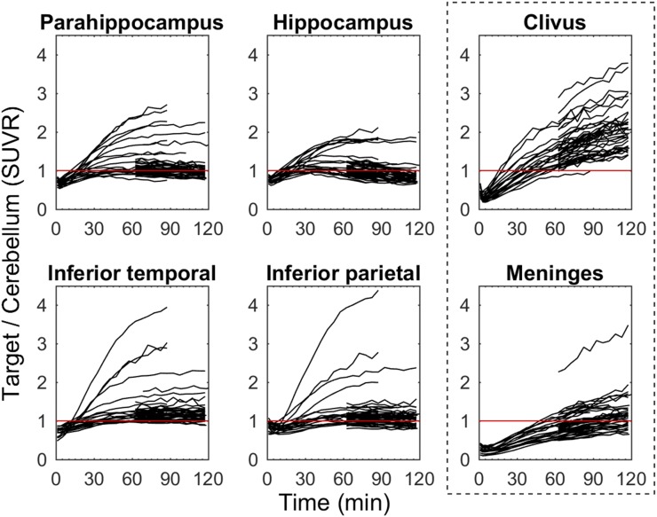
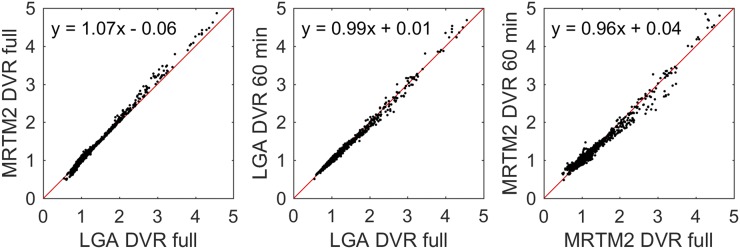
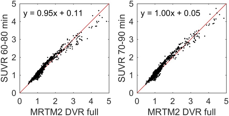
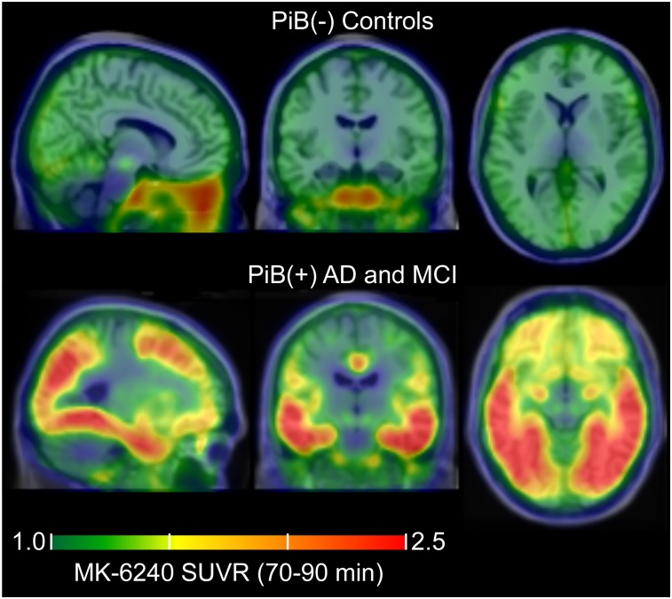
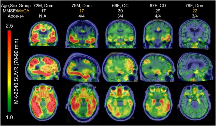
Tau PET imaging has potential for elucidating changes in the deposition of neuropathological tau aggregates that are occurring during the progression of Alzheimer disease (AD). This work investigates in vivo kinetics, quantification strategies, and imaging characteristics of a novel tau PET radioligand 18F-MK-6240 in humans. Methods: Fifty-one individuals ranging from cognitively normal young controls to persons with dementia underwent T1-weighted MRI as well as 11C-PiB and 18F-MK-6240 PET imaging. PET data were coregistered to the MRI, and time-activity curves were extracted from regions of interest to assess 18F-MK-6240 kinetics. The pons and inferior cerebellum were investigated as potential reference regions. Reference tissue methods (Logan graphical analysis [LGA] and multilinear reference tissue method [MRTM2]) were investigated for quantification of 18F-MK-6240 distribution volume ratios (DVRs) in a subset of 19 participants. Stability of DVR methods was evaluated using truncated scan durations. SUV ratio (SUVR) estimates were compared with DVR estimates to determine the optimal timing window for SUVR analysis. Parametric SUVR images were used to identify regions of potential off-target binding and to compare binding patterns with neurofibrillary tau staging established in neuropathology literature. Results: SUVs in the pons and the inferior cerebellum indicated consistent clearance across all 51 subjects. LGA and MRTM2 DVR estimates were similar, with LGA slightly underestimating DVR compared with MRTM2. DVR estimates remained stable when truncating the scan duration to 60 min. SUVR determined 70-90 min after injection of 18F-MK-6240 indicated linearity near unity when compared with DVR estimates and minimized potential spill-in from uptake outside the brain. 18F-MK-6240 binding patterns in target regions were consistent with neuropathological neurofibrillary tau staging. Off-target binding regions included the ethmoid sinus, clivus, meninges, substantia nigra, but not the basal ganglia or choroid plexus. Conclusion:18F-MK-6240 is a promising PET radioligand for in vivo imaging of neurofibrillary tau aggregates in AD with minimal off-target binding in the human brain.
Keywords: Alzheimer’s disease; MK-6240; positron emission tomography; quantification; tau.
© 2019 by the Society of Nuclear Medicine and Molecular Imaging.
Figures
References
-
- Braak H, Braak E. Neuropathological stageing of Alzheimer-related changes. Acta Neuropathol (Berl). 1991;82:239–259. - PubMed
-
- Thal DR, Rüb U, Orantes M, Braak H. Phases of Aβ-deposition in the human brain and its relevance for the development of AD. Neurology. 2002;58:1791–1800. - PubMed
-
- Braak H, Thal DR, Ghebremedhin E, Del Tredici K. Stages of the pathologic process in Alzheimer disease: age categories from 1 to 100 years. J Neuropathol Exp Neurol. 2011;70:960–969. - PubMed
Publication types
MeSH terms
Substances
Grants and funding
LinkOut - more resources
Full Text Sources
Other Literature Sources
Medical