. 2018 Jun 28;131(12):jcs212647.
doi: 10.1242/jcs.212647.

Septin-dependent remodeling of cortical microtubule drives cell reshaping during epithelial wound healing

Affiliations

Septin-dependent remodeling of cortical microtubule drives cell reshaping during epithelial wound healing

Asako Shindo et al. J Cell Sci. .

Abstract

Wounds in embryos heal rapidly through contraction of the wound edges. Despite well-recognized significance of the actomyosin purse string for wound closure, roles for other cytoskeletal components are largely unknown. Here, we report that the septin cytoskeleton cooperates with actomyosin and microtubules to coordinate circumferential contraction of the wound margin and concentric elongation of wound-proximal cells in Xenopus laevis embryos. Microtubules reoriented radially, forming bundles along lateral cell cortices in elongating wound-proximal cells. Depletion of septin 7 (Sept7) slowed wound closure by attenuating the wound edge contraction and cell elongation. ROCK/Rho-kinase inhibitor-mediated suppression of actomyosin contractility enhanced the Sept7 phenotype, whereas the Sept7 depletion did not affect the accumulation of actomyosin at the wound edge. The cortical microtubule bundles were reduced in wound-proximal cells in Sept7 knockdown (Sept7-KD) embryos, but forced bundling of microtubules mediated by the microtubule-stabilizing protein Map7 did not rescue the Sept7-KD phenotype. Nocodazole-mediated microtubule depolymerization enhanced the Sept7-KD phenotype, suggesting that Sept7 is required for microtubule reorganization during cell elongation. Our findings indicate that septins are required for the rapid wound closure by facilitating cortical microtubule reorganization and the concentric elongation of surrounding cells.

Keywords: Cytoskeleton; Embryonic wound healing; Epidermis; Xenopus laevis.


Conflict of interest statement

Competing interestsThe authors declare no competing or financial interests.

Figures
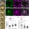
Fig. 1.
Fig. 1.
Cells elongate radially toward the center of the wound. (A) Stereoscope images of the wound made in the epidermal tissue of X. laevis neurula; stills were taken every 2 min from time-lapse movies. Only the outer layer was peeled off. The brown sheet is the outer layer of the epidermis and the paler region shows the exposed deeper layer. The wound edge is surrounded by a dotted line. (B,C) Fluorescence images of wounded embryos injected with membrane-BFP (green) and Lifeact-RFP (F-actin, magenta). Images in B were taken at 3 min after wounding, those in C at 18 min after wounding. Dotted lines surround the wound edge, arrows indicate F-actin accumulation at the wound margin. (D) Quantification of cell elongation during live imaging. The cell elongation index was calculated according to the ellipticity (length:width ratio) of each cell. The x-axis indicates the time after wounding. 5 min (n=27), 10 min (n=33), 20 min (n=39) from 4 embryos. (****P<0.0001, **P=0.0037, one-way ANOVA was applied, followed by the Kruskal–Wallis test for multiple comparisons). (E) Quantification of the length of the cell edge facing the wound. The x-axis indicates the time after wounding. 5 min (n=27), 10 min (n=32), 20 min (n=39) from 4 embryos. (****P<0.0001, **P=0.0013, one-way ANOVA was applied, followed by the Kruskal–Wallis test for multiple comparisons). Scale bars: 20 µm.
Fig. 2.
Fig. 2.
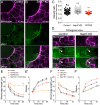
Microtubules are required for cell elongation during wound closure. (A–C′) Still fluorescence images from time-lapse imaging of control (A,A′), nocodazole- (B,B′) or Y27632-treated embryos (C,C′) at 3 min (A,B,C) or 15 min (A′,B′,C′) after wounding. Embryos express membrane-BFP. Scale bars: 20 µm. Dotted lines indicate the wound edge. (D) Quantification of cell elongation by measuring the ellipticity of each cell. (P<0.0001, two-way ANOVA followed by Dunnett's test for multiple comparisons. Control: n=68 from 3 embryos; nocodazole: n=74 from 4 embryos; Y27632: n=73 from 4 embryos; nocodazole+Y27632: n=50 from 3 embryos). (E) Quantification of wound edge shortening of each cell. Control (black), nocodazole-treated (blue), Y27632-treated (red), and mixture of nocodazole and Y27632-treated embryos (purple). (P<0.0001, two-way ANOVA followed by Dunnett's test for multiple comparisons. Control: n=51 from 3 embryos; nocodazole: n=70 from 4 embryos; Y27632: n=78 from 4 embryos; nocodazole+Y27632: n=41 from 3 embryos). (F) Schematic of wound edge and lateral edge of each cell as measured in G–I. (G) Comparison of avarage wound edge length in each cell at 3 minutes after wounding. (***P=0.0002, Control (Cont): n=48 from 3 embryos, nocodazole (Noco): n=62 from 4 embryos, Y27632 (Y27): n=87 from 4 embryos). (H,I) Comparison of average velocity of wound edge shortening (H) or lateral edge elongation (I) at 3–10 min after wounding. (**P=0.0054, ***P<0.0001, one-way ANOVA followed by the Kruskal–Wallis test for multiple comparisons, Cont: n=45 from 3 embryos, Noco: n=72 from 4 embryos, Y27: n=79 from 4 embryos).
Fig. 3.
Fig. 3.
Microtubules reorient during wound closure. (A–C′) Fluorescence images of maximum intensity z-projections of 10-µm thick intact epidermis (A), 5 min after wounding (B) and 10 min after wounding (C) in X. laevis neurula injected with α-tubulin-emerald GFP (green) and membrane RFP (magenta). Dotted arrowheads indicate lack of filaments. Arrowheads indicate microtubule filaments along the lateral cell cortices. A′,B′,C′ are magnified images of single cell–cell junctions (boxed areas in A,B,C, respectively). Dotted lines indicates the wound edge. (D) Microtubule filament formation along cell cortices was quantified by the ratio of α-tubulin-emerald GFP and membrane-RFP. The mean intensity of each marker was measured along the cell–cell junctions with 1-µm width using ImageJ (****P<0.0001, Student’s t-test, Intact: n=40 from 3 embryos, Wounded n=32 from 2 embryos). (E) Same analysis as shown in D but of fixed embryos stained with α-tubulin and β-catenin antibodies shown in Fig. S3. (****P<0.0001, Mann–Whitney U-test, n=90 from 3 embryos, Wounded n=90 from 2 embryos). Random sampling was performed by using software from the free software environment R (https://www.r-project.org/); original number was 241 (intact) and 98 (wounded). Scale bars: 20 µm.
Fig. 4.
Fig. 4.
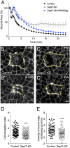
Sept7 is required for rapid wound closure. (A) Quantification of wound area reduction in control, Sept7-KD and sept7 mRNA-expressing embryos. The wound area was plotted every 20 s during the first 4 min, and every minute from 4 min onwards after wounding for each group. Control embryos (n=11), Sept7-KD embryos (n=13, 35 ng MO per blastomere), Sept7-KD embryos injected with sept7 mRNA (80 pg) (n=14) (P<0.0001, two-way ANOVA). (B–C′) Still images from time-lapse imaging of membrane-RFP during wound closure in control embryos (B,B′) and Sept7-KD embryos (C,C′); at 2 min, and at 5 min after wounding. Dotted lines indicate the wound edge. (D) Quantification of cell elongation by measuring ellipticity of each cell surrounding the wound at 15 min after wounding. Control (n=138 from 4 embryos), Sept7-KD embryos (n=136 from 4 embryos). (E) Velocity of cell edge shortening at the wound edge from 2–5 min after wounding in the control (n=53 from 3 embryos) and Sept7-KD embryos (n=71 from 4 embryos). ****P<0.0001, Mann–Whitney U-test. Scale bar: 20 µm.
Fig. 5.
Fig. 5.
Actomyosin requires Sept7 for function but not for formation at the wound edge. (A,B) z-Projection of the immunostained wound area. Control (A) and Sept7-KD (B) embryos were injected with membrane-GFP and stained with anti-pMLC and anti-GFP antibodies. (C) Quantification of pMLC accumulation at each cell junction along the wound edge, normalized to the mean intensity of lateral cell edge in a cell (Control: n=76 from 3 embryos; Sept7-KD: n=125 from 4 embryos; Y27632: n=52 from 3 embryos). One-way ANOVA followed by the Kruskal–Wallis test for multiple comparisons (****P<0.0001). (D) Orthogonal view of the z-projection created by using ImageJ. Dotted lines indicate the outer layer closing the wound. Arrowheads indicate the pMLC ring at the wound edge. (E,E′) Quantification of cell edge length at the wound margin and ellipticity of each cell as an index of cell elongation (ellipticity). Two-way ANOVA was followed by Dunnett's test for multiple comparisons. Control: n=68 from 3 embryos; Y27632 (low): 20 µM, n=67 from 3 embryos; Sept7-KD (low): 17.5 ng MO, n=57 from 3 embryos; Sept7-KD+Y27632 (low): n=54 from 3 embryos. (F,F′) Same analyses as E and E’. Two-way ANOVA was followed by Tukey's test for multiple comparisons. Control: n=65 from 3 embryos; Calyculin A: 125 nM, n=65 from 3 embryos; Sept7-KD: 35 ng MO, n=61 from 3 embryos; Sept7-KD+Calyculin A: n=90 from 4 embryos. Scale bars: 20 µm.
Fig. 6.
Fig. 6.
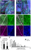
Sept7 coordinates microtubule reorientation after wounding. (A–D) Immunostaining of α-tubulin (green) for microtubules and GFP (magenta) for the membrane. Control or Sept7-KD embryos were fixed 15 min after wounding. Dotted lines indicate the wounded edge; boxed areas in A and C are shown magnified in B and D, respectively; scale bars: 20 µm. (E) Pixel intensities of α-tubulin and the membrane measured on the yellow line drawn across the vertical cell membrane. Pixel intensities were used to create the profile plot in F. (F) Patterns of profile plots of α-tubulin and the membrane. Single, double or triple peaks of α-tubulin reflect cohesiveness of microtubule filaments and the cell membrane. (G) Relative frequency of the three patterns of profile plots shown in F. (H) Histogram showing the distribution of the distance between the peaks (interpeak distance) of α-tubulin and membrane in the profile plots. Control: n=367; KD: n=171; n=2 embryos per group. Range of each bin is 0.16 µm. Data were analyzed using the Kolmogorov-Smirnov test (P<0.0001).
Fig. 7.
Fig. 7.
Sept7 controls microtubule reorientation independently from microtubule stabilization. (A–D) Immunostaining of acetylated α-tubulin (Ac-α-tubulin) in stabilized microtubules, of β-catenin in the cell–cell junction and of GFP in cells overexpressing Map7-GFP. Images in A and B are tile scanned, boxed areas in A and B are shown magnified in C and D, respectively; scale bars: 20 µm (A,B), 10 µm (C,D). (E) Relative frequency of three patterns of plot profiles of Ac-α-tubulin. Analyses are the same as those described for Fig. 4E–G. (F) Histogram showing the distribution of the distance between the intensity peaks of Ac-α-tubulin and β-catenin in the profile plot. Control: n=87; KD: n=90, from 2 embryos per group. Range of each bin is 0.16 µm. Data was analyzed using the Kolmogorov-Smirnov test (P<0.0001).
Fig. 8.
Fig. 8.
Sept7 and microtubules functionally interact during wound closure. (A–D) Live imaging of cell shape in the wounded epidermis of the embryos at 15 min after wounding. Embryos were co-injected with membrane-BFP and α-tubulin-emerald GFP to confirm the effects of nocodazole (Noco). Images show only membrane BFP (white). Embryos were treated with DMSO as control (A), a low concentration (5 µM) of nocodazole (B), weak knockdown of Sept7 (17.5 ng MO) (C) and the combination of weak Sept7 knockdown and low concentration of nocodazole (D). Dotted lines indicate the wound edge. Scale bars: 20 µm. (E) Quantification of cell elongation by measuring the ellipticity of each cell attached to the wound. *P=0.0444, ****P<0.0001, two-way ANOVA followed by Dunnett's test for multiple comparisons. Control: n=60 from 3 embryos; Noco (5 µM): n=74 from 4 embryos; Sept7-KD (17.5 ng of MO): n=57 from 4 embryos; KD+Noco (17.5 ng MO+5 µM nocodazole): n=71 from 4 embryos. (F) Average comparison of wound edge length in each cell at 3 minutes after wounding. *P=0.0284 (nocodazole versus nocodazole+Sept7-MO). *P=0.0421 (Sept7-KD versus nocodazole+Sept7-KD). One-way ANOVA followed by Kruskal–Wallis test for multiple comparisons. (Control: n=39 from 3 embryos, nocodazole: n=54 from 3 embryos, sept7-MO: n=49 from 3 embryos, Sept7-KD+nocodazole: n=54 from 3 embryos). (G,H) Average comparisons of velocity of wound edge shortening (G) or lateral edge elongation (H) at 3–10 min after wounding. *P=0.0468, ***P<0.0001; one-way ANOVA followed by Kruskal–Wallis test for multiple comparisons, (Control: n=35 from 3 embryos, nocodazole: n=42 from 3 embryos, Sept7-KD: n=38 from 3 embryos, Sept7-KD+nocodazole: n=58 from 4 embryos).

References

    1. Abreu-Blanco M. T., Verboon J. M. and Parkhurst S. M. (2011). Cell wound repair in Drosophila occurs through three distinct phases of membrane and cytoskeletal remodeling. J. Cell Biol. 193, 455-464. 10.1083/jcb.201011018 - DOI - PMC - PubMed
    1. Abreu-Blanco M. T., Verboon J. M., Liu R., Watts J. J. and Parkhurst S. M. (2012). Drosophila embryos close epithelial wounds using a combination of cellular protrusions and an actomyosin purse string. J. Cell Sci. 125, 5984-5997. 10.1242/jcs.109066 - DOI - PMC - PubMed
    1. Ageta-Ishihara N., Miyata T., Ohshima C., Watanabe M., Sato Y., Hamamura Y., Higashiyama T., Mazitschek R., Bito H. and Kinoshita M. (2013). Septins promote dendrite and axon development by negatively regulating microtubule stability via HDAC6-mediated deacetylation. Nat. Commun. 4, 2532 10.1038/ncomms3532 - DOI - PMC - PubMed
    1. Bai X., Bowen J. R., Knox T. K., Zhou K., Pendziwiat M., Kuhlenbäumer G., Sindelar C. V. and Spiliotis E. T. (2013). Novel septin 9 repeat motifs altered in neuralgic amyotrophy bind and bundle microtubules. J. Cell Biol. 203, 895-905. 10.1083/jcb.201308068 - DOI - PMC - PubMed
    1. Bement W. M., Mandato C. A. and Kirsch M. N. (1999). Wound-induced assembly and closure of an actomyosin purse string in Xenopus oocytes. Curr. Biol. 9, 579-587. 10.1016/S0960-9822(99)80261-9 - DOI - PubMed

Publication types

LinkOut - more resources