. 2018 Jul 13;293(28):10949-10962.
doi: 10.1074/jbc.RA117.001569. Epub 2018 May 18.

Sequence diversity of tubulin isotypes in regulation of the mitochondrial voltage-dependent anion channel

Affiliations

Sequence diversity of tubulin isotypes in regulation of the mitochondrial voltage-dependent anion channel

Tatiana K Rostovtseva et al. J Biol Chem. .

Abstract

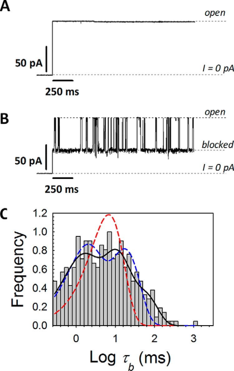
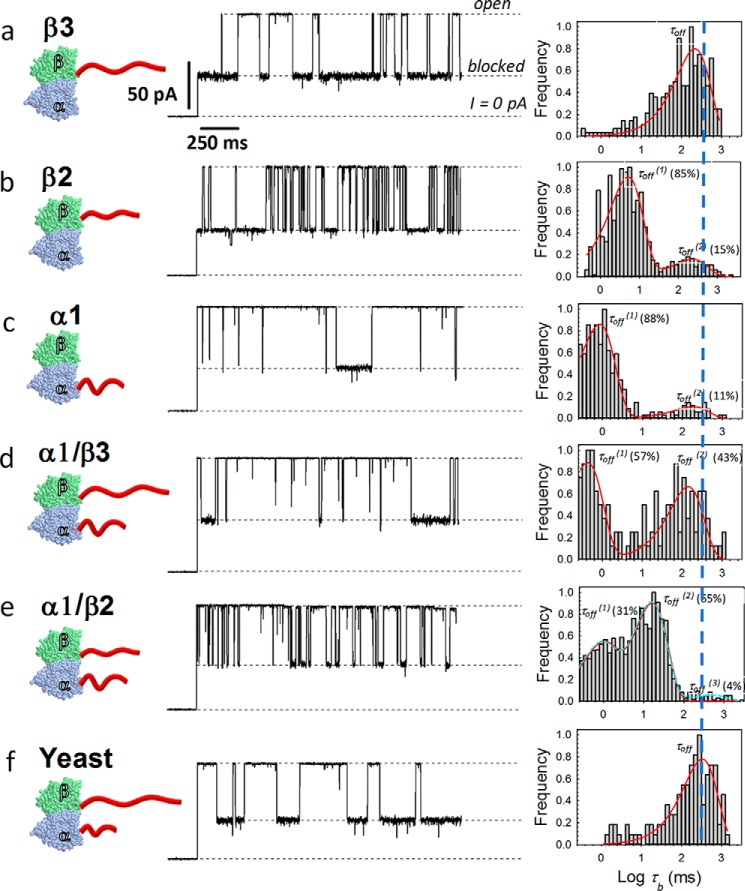
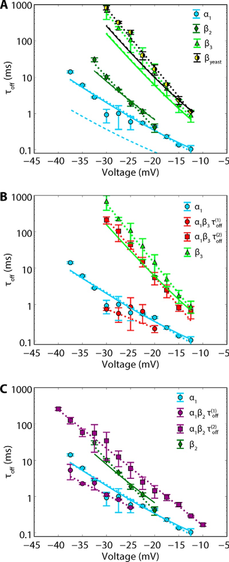
The microtubule protein tubulin is a heterodimer comprising α/β subunits, in which each subunit features multiple isotypes in vertebrates. For example, seven α-tubulin and eight β-tubulin isotypes in the human tubulin gene family vary mostly in the length and primary sequence of the disordered anionic carboxyl-terminal tails (CTTs). The biological reason for such sequence diversity remains a topic of vigorous enquiry. Here, we demonstrate that it may be a key feature of tubulin's role in regulation of the permeability of the mitochondrial outer membrane voltage-dependent anion channel (VDAC). Using recombinant yeast α/β-tubulin constructs with α-CTTs, β-CTTs, or both from various human tubulin isotypes, we probed their interactions with VDAC reconstituted into planar lipid bilayers. A comparative study of the blockage kinetics revealed that either α-CTTs or β-CTTs block the VDAC pore and that the efficiency of blockage by individual CTTs spans 2 orders of magnitude, depending on the CTT isotype. β-Tubulin constructs, notably β3, blocked VDAC most effectively. We quantitatively described these experimental results using a physical model that accounted only for the number and distribution of charges in the CTT, and not for the interactions between specific residues on the CTT and VDAC pore. Based on these results, we speculate that the effectiveness of VDAC regulation by tubulin depends on the predominant tubulin isotype in a cell. Consequently, the fluxes of ATP/ADP through the channel could vary significantly, depending on the isotype, thus suggesting an intriguing link between VDAC regulation and the diversity of tubulin isotypes present in vertebrates.

Keywords: C-terminal tail; VDAC; beta-barrel channel; drift-diffusion model; engineered recombinant tubulin; gating; gating charge; intrinsically disordered protein domains; ion channel; membrane protein; membrane transport; mitochondrial transport; peripheral membrane proteins; permeability; protein engineering; protein-lipid interaction; recombinant protein expression; tubulin.


Conflict of interest statement

The authors declare that they have no conflicts of interest with the contents of this article

Figures

Figure 1.
Figure 1.
Mammalian tubulin induces VDAC blockage events characterized by a wide distribution of blockage (dwell) times. A and B, representative records of the current through the same single VDAC channel before (A) and after (B) the addition of 20 nm WT mammalian tubulin (isolated from porcine brain), at −27.5 mV of applied voltage. The membrane-bathing solutions contained 1 m KCl buffered with 5 mm HEPES at pH 7.4. Here and in Fig. 3, dashed lines indicate VDAC open and tubulin-blocked states and zero current. For clarity of presentation, the current records were inverted and smoothed with a 33-point boxcar digital filter using pClamp version 10.3. C, log-binned distribution of blockage times, τb, fit by a single exponent (dashed red line) or a sum of two exponents (dashed blue line). The maximum statistically significant number of exponential components (p < 1 × 10⁻⁵ using χ2 test) is three (solid black line), using a Simplex optimization algorithm and maximum likelihood minimization. The total number of events in the histogram is 459. The position of the left vertical axis corresponds to the time resolution of experimental setup, which is about 0.2 ms.
Figure 2.
Figure 2.
β3 tubulin is found in close proximity to VDAC in neuroblastoma cells. A, representative image of Duolink in situ proximity ligation assay using anti-β3 tubulin and anti-VDAC antibodies in human neuroblastoma cells. Interaction between molecules is indicated by a red positive reaction. B, no reaction was detected in the negative control, in which no primary antibodies were added. Scale bar, 50 μm. Nuclei are visualized using DAPI.
Figure 3.
Figure 3.
Kinetic analysis of VDAC blockage by individual tubulin constructs. Left column, a–f, cartoons illustrating recombinant tubulins with human β3, β2, α1, α1/β3, α1/β2, and yeast CTTs (red). Middle column, representative traces of ion currents through a single VDAC channel in the presence of 8–10 nm recombinant tubulin construct shown at the left. All records were taken at −27.5 mV of applied voltage; constructs were added to the cis compartment. Other experimental conditions are as in Fig. 1. Right column, corresponding log-binned distributions of blockage times, τb, calculated from statistical analysis of current records, fragments of which are shown in the middle. Red solid lines are the single exponential fits with characteristic time τoff = 〈τb〉 equal to 242 and 330 ms for β3 and yeast tubulin WT (a and f) or double exponential fits with characteristic times τoff(1) and τoff(2) equal to 5 and 229 ms for β2 (b), 0.9 and 184 ms for α1 (c), 0.4 and 142 ms for α1/β3 (d), and 0.8 and 16 ms for α1/β2 (e). The best fit (p < 1 × 10⁻⁵ using χ2 test) for the α1/β2 construct (e) is achieved with three exponents (dashed cyan line), where the minor third component (τoff(3) = 488 ms) accounts for 4% of events. The occurrence (%) of blockage times in the log-binned distribution of blockage times is shown in brackets for all double and triple exponential fits. The total number of events represented by histograms in a, b, c, d, e, and f are 362, 778, 618, 269, 1488, and 288, respectively.
Figure 4.
Figure 4.
Mean dwell times of tubulin blockage are exponentially voltage-dependent. A, voltage dependences of τoff(1) obtained for α1, β2, and β3 tubulin constructs and βyeast. B and C, voltage dependences of τoff(1) and τoff(2) obtained for the α1/β3 construct compared with β3 and α1 constructs (B) and for the α1/β2 construct compared with β2 and α1 constructs (C). Dotted lines, fits to the Arrhenius equation (Equation 1); solid lines, fits to the drift-diffusion model (see “Appendix”) with optimized parameters ltα = 1.43 nm and ltβ = 2.01 nm; dashed lines are initial estimates using ltα = ltβ = 2 nm as reasonable values of the drift-diffusion model. Each data point is a mean of 3–5 independent experiments ± S.D. (error bars).
Figure 5.
Figure 5.
Ionic selectivity of the tubulin-blocked state of VDAC is cationic for both a recombinant construct and WT mammalian tubulin. Current–voltage relationships for a single VDAC channel in a membrane separating 200 mm (cis) and 1 m (trans) KCl solutions (2 mm HEPES, pH 7.4) are shown. Open state (black triangles) has anionic selectivity with the reversal potential ψrev = −9.4 ± 0.5 mV, marked by the black arrow. Currents for the tubulin-blocked states are denoted by red circles for the α1/β3 tubulin construct and by blue circles for WT mammalian tubulin. To be able to measure the reversal potential for the two tubulins on the same single channel, we added the recombinant tubulin to the cis side and WT tubulin to the trans side of the membrane, in concentrations of 10 and 20 nm, respectively. As indicated by the arrows, blockage of VDAC by either tubulin similarly reverses the original anionic selectivity of the channel to the cationic one with ψrev = 8.4 ± 0.4 mV and 9.5 ± 0.6 mV for α1/β3 and the WT mammalian tubulin, respectively.
Figure 6.
Figure 6.
The on-rate constants of VDAC blockage by different tubulin constructs. There is no significant difference between the on-rate constants of VDAC blockages except for the difference between β2 and α1/β2 (*, p < 0.05 one-way ANOVA). Data were analyzed by one-way ANOVA (F4,14 = 5.33, p = 0.01) followed by a pairwise multiple Holm–Sidak comparison test. Applied voltage was −25 mV. Each symbol corresponds to an individual experiment; bars are the mean values of 3–5 independent experiments ± S.D. (error bars). When the on-rate constants are normalized by the number of CTTs per construct, the differences between β2 and α1/β2 lose statistical significance (F4,14 = 1.29, p = 0.32, one-way ANOVA).
Figure 7.
Figure 7.
Physical model of CTT/pore interaction. A, geometry of the model showing the definitions of pore length lp and tethering distance lt. B, potentials corresponding to the escape of each CTT from the pore, calculated using optimized parameters ltα = 1.43 nm and ltβ = 2.01 nm. Differences in escape times are described almost entirely by the heights of the barrier and not by residue-specific interactions between the CTT and the pore lumen.
Figure 8.
Figure 8.
Cartoon of a model of VDAC pore blockage by the α-CTT or β-CTT of tubulin. The orientation of membrane-bound tubulin dimer is adopted from Ref. with α-tubulin as the membrane-bound subunit. In such alignment, the contribution of the length and especially the first amino acids of the CTT to the blocking kinetics should be different for α-CTTs (a) and β-CTTs (b). There is some degree of rotational dynamics for the stably bound α-tubulin shown in (b) with the arrow and shadow image, which could also affect the kinetics of pore blockage by the β-CTT.

References

    1. Nogales E., Wolf S. G., and Downing K. H. (1998) Structure of the αβ tubulin dimer by electron crystallography. Nature 391, 199–203 10.1038/34465 - DOI - PubMed
    1. Roll-Mecak A. (2015) Intrinsically disordered tubulin tails: complex tuners of microtubule functions? Semin. Cell Dev. Biol. 37, 11–19 10.1016/j.semcdb.2014.09.026 - DOI - PMC - PubMed
    1. Sirajuddin M., Rice L. M., and Vale R. D. (2014) Regulation of microtubule motors by tubulin isotypes and post-translational modifications. Nat. Cell Biol. 16, 335–344 10.1038/ncb2920 - DOI - PMC - PubMed
    1. Ludueña R. F. (1993) Are tubulin isotypes functionally significant. Mol. Biol. Cell 4, 445–457 10.1091/mbc.4.5.445 - DOI - PMC - PubMed
    1. Redeker V. (2010) Mass spectrometry analysis of C-terminal posttranslational modifications of tubulins. Methods Cell Biol. 95, 77–103 10.1016/S0091-679X(10)95006-1 - DOI - PubMed

Publication types