Case Reports
. 2018 May 18;7(1):93.
doi: 10.1038/s41426-018-0095-0.

The first isolate of Candida auris in China: clinical and biological aspects

Affiliations
Case Reports

The first isolate of Candida auris in China: clinical and biological aspects

Xiaojuan Wang et al. Emerg Microbes Infect. .

Abstract

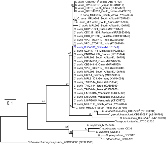
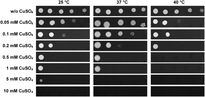
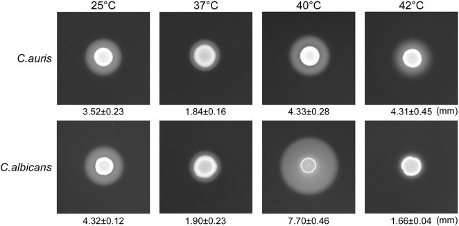
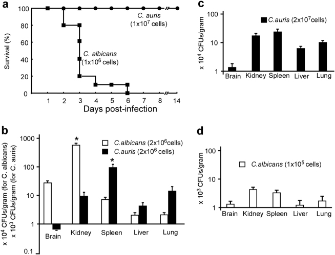
The emerging human fungal pathogen Candida auris has been recognized as a multidrug resistant species and is associated with high mortality. This fungus was first described in Japan in 2009 and has been reported in at least 18 countries on five continents. In this study, we report the first isolate of C. auris from the bronchoalveolar lavage fluid (BALF) of a hospitalized woman in China. Interestingly, this isolate is susceptible to all tested antifungals including amphotericin B, fluconazole, and caspofungin. Copper sulfate (CuSO4) also has a potent inhibitory effect on the growth of this fungus. Under different culture conditions, C. auris exhibits multiple morphological phenotypes including round-to-ovoid, elongated, and pseudohyphal-like forms. High concentrations of sodium chloride induce the formation of a pseudohyphal-like form. We further demonstrate that C. auris is much less virulent than Candida albicans in both mouse systemic and invertebrate Galleria mellonella models.


Conflict of interest statement

The authors declare that they have no conflict of interest.

Figures

Fig. 1
Fig. 1. Phylogenetic trees generated by the Maximum- Likelihood (ML) method.
Internal transcribed spacer (ITS) sequences of nuclear rDNA of Candida auris, C. auris closely related species, and Schizosaccharomyces pombe were used. The GenBank accession numbers are shown in the brackets. Candida species used: C. duobushaemulonii, C. pseudohaemulonii, C. haemulonis, C. tropicalis, C. dubliniensis, C. albicans, C. parapsilosis, and C. orthopsilosis. The Maximum-Likelihood phylogenetic tree was generated using RAxML based on the General Time Reversible (GTR) model and Gamma distribution with Invariant sites (G + I). The percentages of replicate trees in which the associated taxa clustered together in the bootstrap test (1000 replicates) are indicated at the branches. The scale bar indicates the nucleotide substitutions per site. Strain BJCA001 is highlighted in blue
Fig. 2
Fig. 2. Inhibitory effect of CuSO4 on the growth of C. auris grown at 25 °C, 37 °C, and 40 °C.
C. auris cells were adjusted to 5 × 108 cells/mL, and then 10-fold serial dilutions of cells (2 μL) were spotted onto YPD and YPD containing CuSO4 media for four days of growth
Fig. 3
Fig. 3. Morphologies of C. auris grown on Lee’s glucose, Lee’s GlcNAc, Spider, and agar + serum media.
Cells (1 × 105) were spotted onto different medias and cultured at 25 °C, 37 °C, and 40 °C for five days. Scale bar, 10 μm
Fig. 4
Fig. 4. Morphologies of C. auris cells grown on YPD (a) and YPD plus 10% NaCl (b) media.
Cells (1 × 105) were spotted onto different medias and cultured at 37 °C and 40 °C for five days. Cells were collected and stained with DAPI or Calcofluor white. Scale bar, 10 μm. DIC, differential interference contrast
Fig. 5
Fig. 5. Scanning electron microscopy images (SEM) of C. auris cells grown on YPD and YPD plus 10% NaCl media.
Cells (1 × 105) were spotted onto different media and cultured at 37 °C and 40 °C for five days. Cells were then collected for SEM assays. Scale bar, 5 μm
Fig. 6
Fig. 6. A comparison of Sap activities of C. auris and C. albicans.
We spotted 5 × 106 cells of C. auris or C. albicans (SC5314) in 5 µL ddH2O onto YCB-BSA medium plates, followed by growth at 25 °C, 37 °C, 40 °C, and 42 °C for five days. The white precipitation zones (halos) around the cell spots indicate Sap-mediated BSA hydrolysis. The width of the precipitation zones is indicated below the corresponding image. Average values of three biological repeats and standard deviations are presented
Fig. 7
Fig. 7. Virulence of C. auris and C. albicans in mouse systemic infection models.
a Survival curves of mice injected with C. auris (1 × 107 cells/mouse) and C. albicans (1 × 106 cells/mouse) via the lateral tail vein. Ten mice were used for each strain. b–d Fungal burden assays. Five mice were used for each infection group. Mice were killed for CFU assays at 24 h post-infection. b Each mouse was injected with 2 × 106 cells of C. auris or C. albicans. c Each mouse was injected with 2 × 107 cells of C. auris. d, Each mouse was injected with 1 × 105 cells of C. albicans. * Indicates a significant difference (P vale < 0.01, Student’s t-test, two-tailed) compared with the fungal burden in other organs

References

    1. Pfaller MA, Diekema DJ. Epidemiology of invasive candidiasis: a persistent public health problem. Clin. Microbiol. Rev. 2007;20:133–163. doi: 10.1128/CMR.00029-06. - DOI - PMC - PubMed
    1. Satoh K, et al. Candida auris sp. nov., a novel ascomycetous yeast isolated from the external ear canal of an inpatient in a Japanese hospital. Microbiol. Immunol. 2009;53:41–44. doi: 10.1111/j.1348-0421.2008.00083.x. - DOI - PubMed
    1. Lockhart SR, et al. Simultaneous Emergence of Multidrug-Resistant Candida auris on 3 Continents Confirmed by Whole-Genome Sequencing and Epidemiological Analyses. Clin. Infect. Dis. 2017;64:134–140. doi: 10.1093/cid/ciw691. - DOI - PMC - PubMed
    1. Spivak, E. S. & Hanson, K. E. Candida auris: an Emerging Fungal Pathogen. J. Clin. Microbiol.56, 10.1128/JCM.01588-17 (2018). - PMC - PubMed
    1. Clancy CJ, Nguyen MH. Emergence of Candida auris: An International Call to Arms. Clin. Infect. Dis. 2017;64:141–143. doi: 10.1093/cid/ciw696. - DOI - PubMed

Publication types

MeSH terms