. 2018 May 18;9(1):1989.
doi: 10.1038/s41467-018-04351-0.

Transient metal-centered states mediate isomerization of a photochromic ruthenium-sulfoxide complex

Affiliations

Transient metal-centered states mediate isomerization of a photochromic ruthenium-sulfoxide complex

Amy A Cordones et al. Nat Commun. .

Abstract

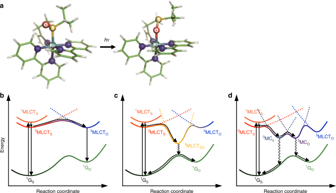
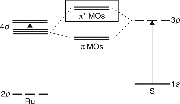
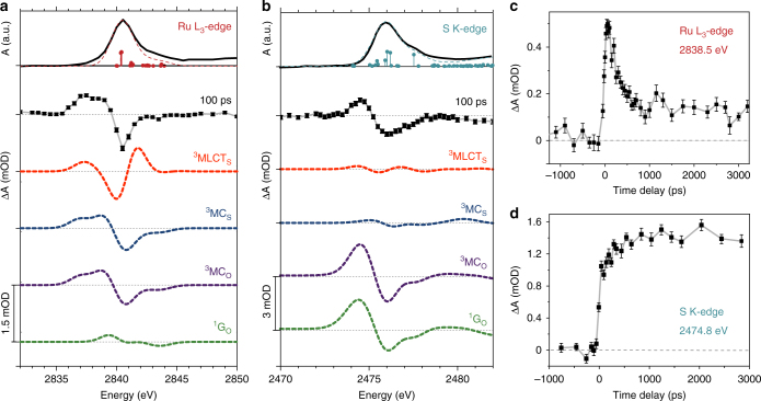
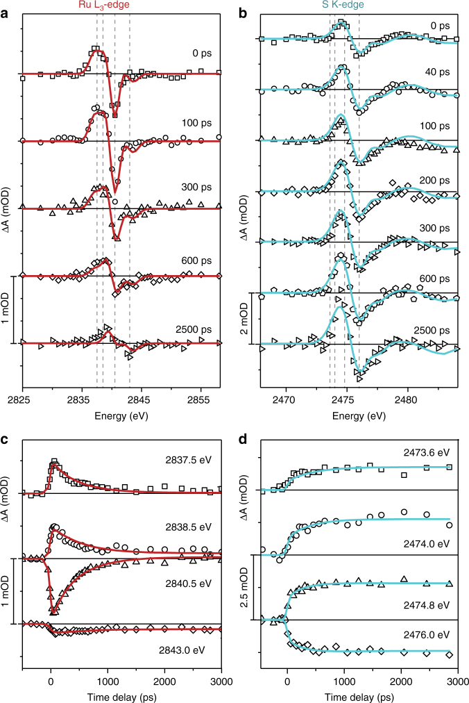
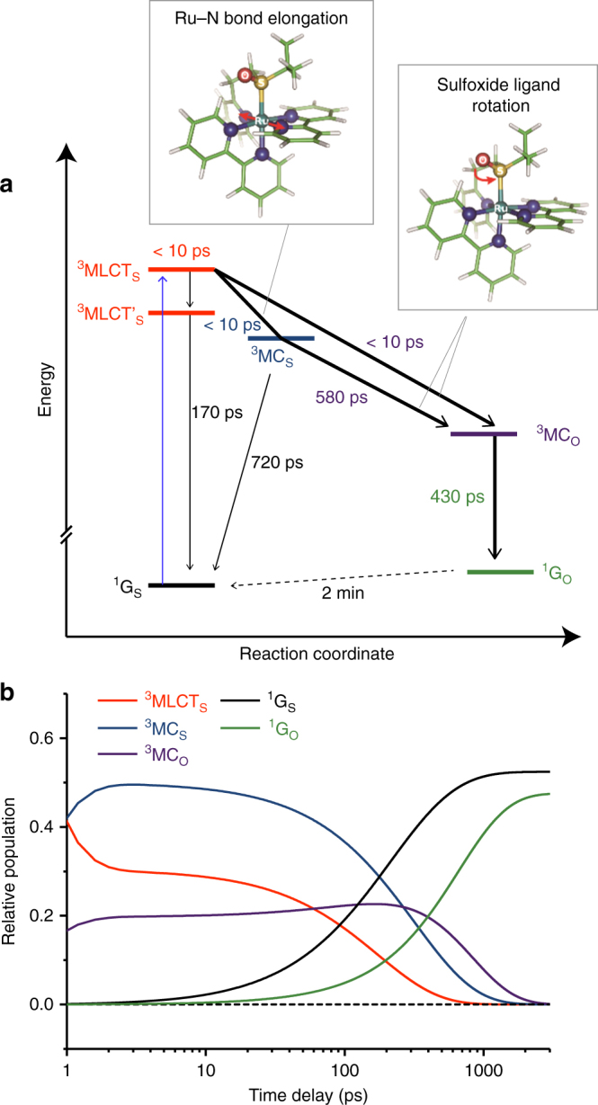
Ultrafast isomerization reactions underpin many processes in (bio)chemical systems and molecular materials. Understanding the coupled evolution of atomic and molecular structure during isomerization is paramount for control and rational design in molecular science. Here we report transient X-ray absorption studies of the photo-induced linkage isomerization of a Ru-based photochromic molecule. X-ray spectra reveal the spin and valence charge of the Ru atom and provide experimental evidence that metal-centered excited states mediate isomerization. Complementary X-ray spectra of the functional ligand S atoms probe the nuclear structural rearrangements, highlighting the formation of two metal-centered states with different metal-ligand bonding. These results address an essential open question regarding the relative roles of transient charge-transfer and metal-centered states in mediating photoisomerization. Global temporal and spectral data analysis combined with time-dependent density functional theory reveals a complex mechanism for photoisomerization with atomic details of the transient molecular and electronic structure not accessible by other means.


Conflict of interest statement

The authors declare no competing interests.

Figures

Fig. 1
Fig. 1
Structures of [Ru(bpy)2(pyESO)]2+ and various photoisomerization pathways. a Molecular structures before and after photoinduced isomerization. Color code: cyan Ru, yellow S, red O, blue N, green C, white H. b Adiabatic photo-isomerization on lowest 3MLCT excited states. c Non-adiabatic photo-isomerization on lowest 3MLCT excited states. d Non-adiabatic photo-isomerization involving 3MLCT and 3MC excited states. Vertical arrows represent photo-absorption and emission processes. Wavy arrows represent non-radiative transitions (conical intersections are not shown for simplicity). Curvy arrows represent adiabatic relaxation paths on potential energy surfaces. Dotted lines represent diabatic states for triplet states. 1GS: S-bonded ground state reactant, 1GO: O-bonded ground state photoproduct, 3MLCTS: S-bonded 3MLCT excited state, 3MLCTO: O-bonded 3MLCT excited state, 3MLCTSO: sideways SO-bonded 3MLCT excited state, 3MCS: S-bonded 3MC excited state, 3MCO: O-bonded 3MC excited state
Fig. 2
Fig. 2
Relevant atomic orbitals probed by XAS experiments. XAS at the Ru L-edge is characterized by transitions between Ru 2p orbitals and molecular orbitals containing primarily Ru 4d character. XAS at the S K-edge is characterized by transitions between S 1s orbitals and molecular orbitals that are an antibonding combination of S 3p and Ru 4d orbitals (“π* MOs” indicated by solid box) and higher-energy ligand-centered orbitals
Fig. 3
Fig. 3
Results of TR-XAS measurements and comparison with TD-DFT simulated spectra. Measured and simulated data at the Ru L3-edge (a) and S K-edge (b). Top panel (solid black trace) is the measured absorption spectrum of [Ru(bpy)2(pyESO)](PF6)2 in the S-bonded ground state. The TD-DFT predicted transitions are indicated as sticks and the predicted spectrum is shown by the dashed line (overlaid with the measured spectrum). Below (second from top, square points) is the measured transient differential absorption spectrum at 100 ps time delay. Error bars indicate the standard error of the mean (obscured in some cases by the large size of the data points). All dashed traces below are TD-DFT simulated differential spectra of several proposed excited state intermediates (S-bonded triplet MLCT and MC states: 3MLCTS and 3MCS; O-bonded MC state: 3MCO) and the O-bonded ground state (1GO). The transient measured and simulated difference spectra are all plotted on the same vertical scale, with the amplitude indicated by the scale bars. c Fixed energy time-delay scan measured at the Ru L3-edge (2838.5 eV), including error bars indicating the standard error of the mean. d Fixed energy time-delay scan measured at the S K-edge (2474.0 eV), with error bars indicating the standard error of the mean
Fig. 4
Fig. 4
Complete TR-XAS data measured for [Ru(bpy)2(pyESO)]2+ and global fit results. Measured TR-XAS data (scattered) is overlaid with the least-square fit to the kinetic model described in the text (solid lines). a, b Differential absorption spectra at several fixed time-delays at the Ru L3-edge (a) and S K-edge (b). Vertical lines indicate the energies monitored as a function of time-delay. c, d Time-delay scans at several Ru L3-edge (c) and S K-edge (d) fixed energies
Fig. 5
Fig. 5
Proposed mechanism and kinetics for the photoisomerization of [Ru(bpy)2(pyESO)]2+. a Proposed photoisomerization mechanism with labels indicating electronic state and lifetime for each ground state and intermediate structure. Bold arrows indicate the basic mechanistic requirements required to describe the Ru and S edge data, including the necessity of MC excited states (Ru edge) and of two isomerization pathways (S edge), and the formation time for the O-bonded ground state. Insets show the major structural changes that accompany electronic transitions. b Time-dependent fractional population for each reaction intermediate

References

    1. Schoenlein RW, Peteanu LA, Mathies RA, Shank CV. The first step in vision: femtosecond isomerization of rhodopsin. Science. 1991;254:412. doi: 10.1126/science.1925597. - DOI - PubMed
    1. Polli D, et al. Conical intersection dynamics of the primary photoisomerization event in vision. Nature. 2010;467:440–443. doi: 10.1038/nature09346. - DOI - PubMed
    1. Saha S, Stoddart JF. Photo-driven molecular devices. Chem. Soc. Rev. 2007;36:77–92. doi: 10.1039/B607187B. - DOI - PubMed
    1. Bonnet S, Collin JP. Ruthenium-based light-driven molecular machine prototypes: synthesis and properties. Chem. Soc. Rev. 2008;37:1207–1217. doi: 10.1039/b713678c. - DOI - PubMed
    1. Jean-Ruel H, et al. Ring-closing reaction in diarylethene captured by femtosecond electron crystallography. J. Phys. Chem. B. 2013;117:15894–15902. doi: 10.1021/jp409245h. - DOI - PubMed

Publication types

LinkOut - more resources