Review
. 2018 Aug;18(8):500-510.
doi: 10.1038/s41568-018-0016-5.

Artificial intelligence in radiology

Affiliations
Review

Artificial intelligence in radiology

Ahmed Hosny et al. Nat Rev Cancer. 2018 Aug.

Abstract

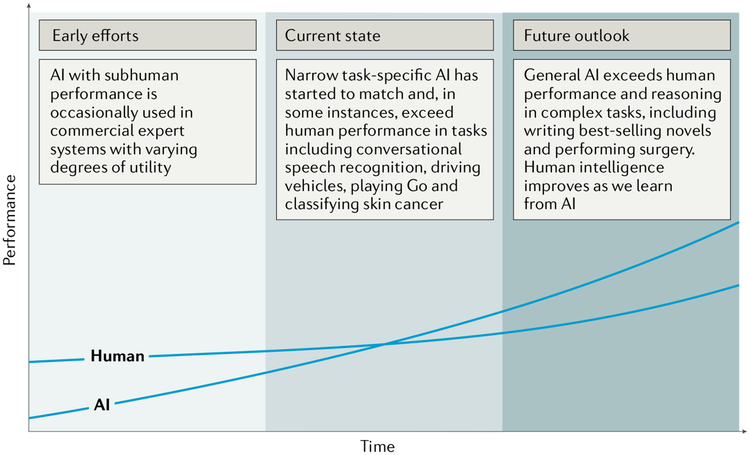
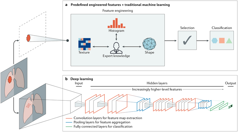
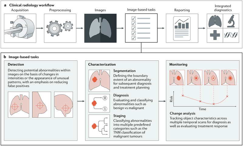
Artificial intelligence (AI) algorithms, particularly deep learning, have demonstrated remarkable progress in image-recognition tasks. Methods ranging from convolutional neural networks to variational autoencoders have found myriad applications in the medical image analysis field, propelling it forward at a rapid pace. Historically, in radiology practice, trained physicians visually assessed medical images for the detection, characterization and monitoring of diseases. AI methods excel at automatically recognizing complex patterns in imaging data and providing quantitative, rather than qualitative, assessments of radiographic characteristics. In this Opinion article, we establish a general understanding of AI methods, particularly those pertaining to image-based tasks. We explore how these methods could impact multiple facets of radiology, with a general focus on applications in oncology, and demonstrate ways in which these methods are advancing the field. Finally, we discuss the challenges facing clinical implementation and provide our perspective on how the domain could be advanced.


Conflict of interest statement

Competing interests

The authors declare no competing interests.

Figures

Fig. 1 |
Fig. 1 |. Artificial versus human intelligence.
This plot outlines the performance levels of artificial intelligence (AI) and human intelligence starting from the early computer age and extrapolating into the future. Early AI came with a subhuman performance and varying degrees of success. Currently, we are witnessing narrow task-specific AI applications that are able to match and occasionally surpass human intelligence–,. It is expected that general AI will surpass human performance in specific applications within the coming years. Humans will potentially benefit from the human-AI interaction, bringing them to higher levels of intelligence.
Fig. 2 |
Fig. 2 |. Artificial intelligence methods in medical imaging.
This schematic outlines two artificial intelligence (AI) methods for a representative classification task, such as the diagnosis of a suspicious object as either benign or malignant. a | The first method relies on engineered features extracted from regions of interest on the basis of expert knowledge. Examples of these features in cancer characterization include tumour volume, shape, texture, intensity and location. The most robust features are selected and fed into machine learning classifiers. b | The second method uses deep learning and does not require region annotation — rather, localization is usually sufficient. It comprises several layers where feature extraction, selection and ultimate classification are performed simultaneously during training. As layers learn increasingly higher-level features (Box 1), earlier layers might learn abstract shapes such as lines and shadows, while other deeper layers might learn entire organs or objects. Both methods fall under radiomics, the data-centric, radiology-based research field.
Fig. 3 |
Fig. 3 |. Artificial intelligence impact areas within oncology imaging.
This schematic outlines the various tasks within radiology where artificial intelligence (AI) implementation is likely to have a large impact. a | The workflow comprises the following steps: preprocessing of images after acquisition, image-based clinical tasks (which usually involve the quantification of features either using engineered features with traditional machine learning or deep learning), reporting results through the generation of textual radiology reports and, finally, the integration of patient information from multiple data sources. b | AI is expected to impact image-based clinical tasks, including the detection of abnormalities; the characterization of objects in images using segmentation, diagnosis and staging; and the monitoring of objects for diagnosis and assessment of treatment response. TNM, tumour–node–metastasis.

References

    1. Editors, N. Auspicious machine learning. Nat. Biomed. Engineer 1, 0036 (2017).
    1. Mnih V et al. Human-level control through deep reinforcement learning. Nature 518, 529–533 (2015). - PubMed
    1. Moravcík M et al. DeepStack: Expert-level artificial intelligence in heads-up no-limit poker. Science 356, 508–513 (2017). - PubMed
    1. Xiong W et al. Toward human parity in conversational speech recognition. IEEE/ACM Trans. Audio Speech Language Process 25, 2410–2423 (2017).
    1. Pendleton SD et al. Perception, planning, control, and coordination for autonomous vehicles. Machines 5, 6 (2017).

Publication types