. 2018 Jul;7(7):3132-3142.
doi: 10.1002/cam4.1485. Epub 2018 May 18.

LGR5 promotes epithelial ovarian cancer proliferation, metastasis, and epithelial-mesenchymal transition through the Notch1 signaling pathway

Affiliations

LGR5 promotes epithelial ovarian cancer proliferation, metastasis, and epithelial-mesenchymal transition through the Notch1 signaling pathway

Wenxue Liu et al. Cancer Med. 2018 Jul.

Abstract

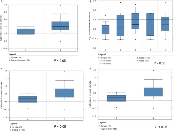
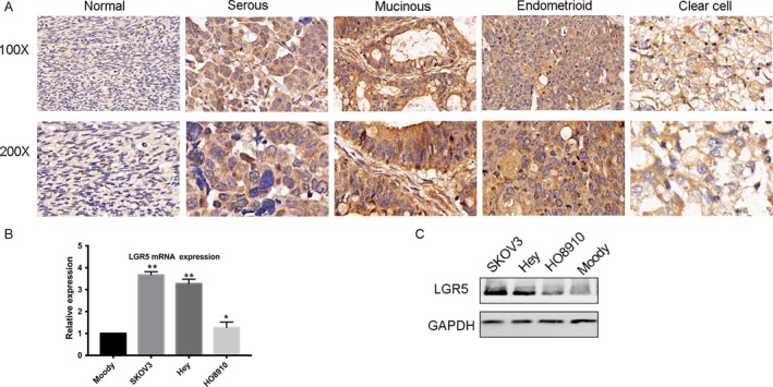
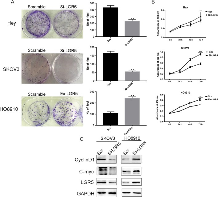
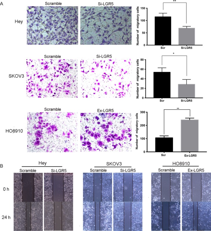
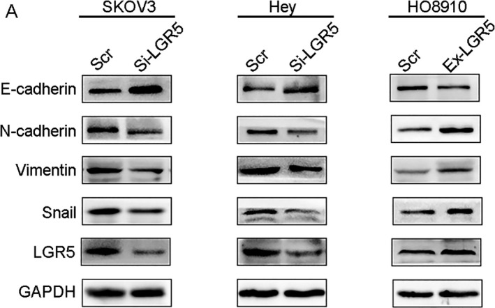
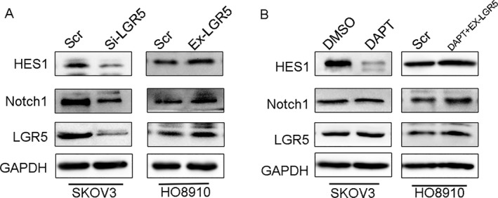
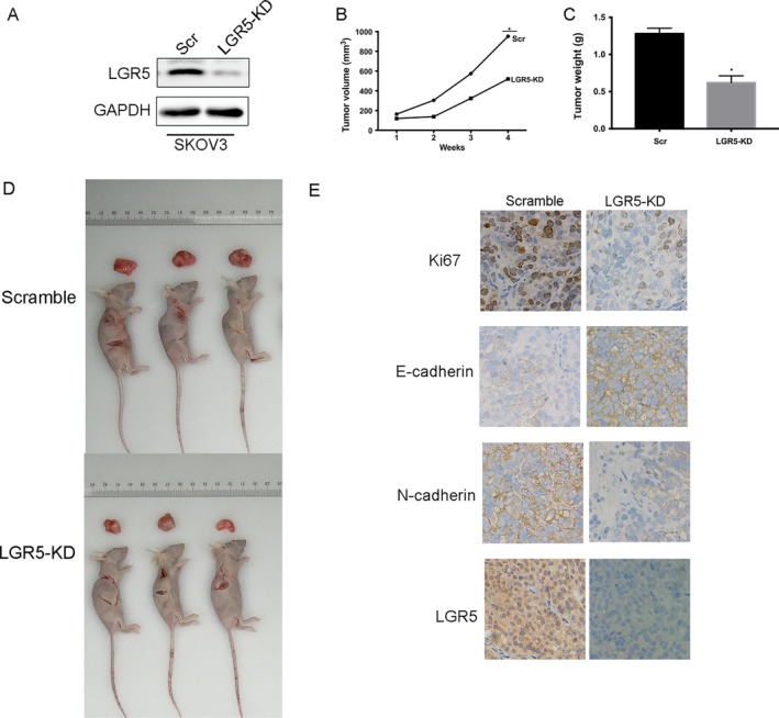
Leucine-rich repeat-containing G protein-coupled receptor 5 (LGR5) plays a vital role in the development of malignant tumors; however, its biological role and underlying mechanism in epithelial ovarian cancer (EOC) remain unclear. In this study, we aimed to investigate the biological function and clinical significance of LGR5 in human EOC. We evaluated LGR5 expression in EOC cell lines and tissues from ovarian cancer patients by qPCR, Western blotting, and immunohistochemical analysis. Cell proliferation, colony formation, transwell invasion assay, and scratch-wound assays were conducted to evaluate the expansion and invasion abilities of EOC cells. Tumor xenograft experiments were performed in female BALB/c athymic nude mice to test cell proliferation in vivo. Western blot analysis was performed to confirm the expression of epithelial-to-mesenchymal transition (EMT) signature proteins and their association with Notch1 signaling. The results demonstrated that LGR5 was overexpressed in EOC tissues and cell lines. Aberrant expression of LGR5 was significantly associated with patient age (P = 0.006), tumor histologic type (P < 0.001), and distant metastasis (P = 0.025). Consistent with these findings, suppression of LGR5 expression led to decreased proliferation and metastasis of EOC cell lines. Furthermore, LGR5 could induce EMT and regulate the Notch1 signaling pathway. Taken together,LGR5 may have an important role in the promotion of tumorigenesis and metastasis of EOC and is a potential therapeutic target for EOC management.

Keywords: Epithelial-mesenchymal transition; LGR5; Notch1 signaling pathway; epithelial ovarian cancer; metastasis.


Figures

Figure 1
Figure 1
Analysis of LGR5 RNA expression based on Oncomine data. (A) LGR5 expression of normal specimens and ovarian carcinoma demonstrating that LGR5 expression levels were increased in ovarian cancer relative to control samples (Cancer Res 2008/07/01). P = 0.0001. (B–C) LGR5 expression levels were increased in grades 1, 2, 3 (Cancer Res 2006/02/01), and 2–3 (Cancer Res 2008/07/01) tumors. P = 0.0001. (D) LGR5 expression levels were increased in stage III or IV tumors compared with normal tissue (Cancer Res 2008/07/01). P = 0.0001.
Figure 2
Figure 2
LGR5 expression and pathologic features in EOC specimens. (A) Immunohistochemical staining of LGR5. LGR5 was overexpressed and localized within the cytoplasm of tumor cells in EOC tissue specimens (scale bar = 20 μm). (B–C) qPCR and Western blot analysis were performed in three EOC cell lines to assess LGR5 expression levels. The human ovarian epithelial cell line, Moody, was used as control.
Figure 3
Figure 3
Elevated expression of LGR5 promotes the proliferation of EOC cells in vitro. (A) LGR5 siRNA (Si‐LGR5) inhibited EOC (Hey and SKOV3 cells) colony formation in vitro, while overexpression of LGR5 in HO8910 cells (Ex‐LGR5) enhanced EOC cell colony formation in contrast with the control (Scramble). (B) Proliferation of Hey, SKOV3, and HO8910 cells treated with LGR5 siRNA (Si‐LGR5) or with LGR5 overexpression (Ex‐LGR5) and their respective controls were evaluated using CCK‐8 assays. (C) Compared to the control group, knockdown of LGR5 (Si‐LGR5) in SKOV3 cells inhibited the expression of cyclin D1 and C‐myc, in contrast to the effects of LGR5 overexpression in HO8910 cells.
Figure 4
Figure 4
LGR5 promotes the migration and invasion capacity of ovarian cancer cells through EMT. (A) Transwell migration assays of Hey and SKOV3 cells treated with LGR5 siRNA (Si‐LGR5), HO8910 cells overexpressing LGR5 (Ex‐LGR5), and their respective controls. Quantification of cells that migrated through the membrane (right) was performed using data from three randomly selected fields of view. Original magnification 200×. Data are presented as means ± SD. *P < 0.05, **P < 0.01, compared with the control group. (B) Wound‐healing migration assays of Hey and SKOV3 cells treated with LGR5 siRNA (Si‐LGR5), HO8910 cells overexpressing LGR5 (Ex‐LGR5), and their respective controls (Scramble). Knockdown of LGR5 in SKOV3 and Hey cells resulted in reduced wound‐healing ability, compared with control cells, which was restored by increase in LGR5 expression in HO8910 cells. Original magnification 200×, scale bar = 20 μm.
Figure 5
Figure 5
Effect of LRG5 on expression of epithelial‐to‐mesenchymal transition markers. (A) Western blots confirmed that E‐cadherin was upregulated while N‐cadherin, Vimentin, and Snail were downregulated in Hey and SKOV3 cells treated with LGR5 siRNA (Si‐LGR5), in contrast with HO8910 cells overexpressing LGR5 (Ex‐LGR5).
Figure 6
Figure 6
LGR5 can activate the Notch1 signaling pathway. (A) Western blot analysis showed that silencing of LGR5 (Si‐LGR5) expression in SKOV3 cells decreased Notch1 expression levels, while increased LGR5 expression (Ex‐LGR5) in HO8910 cells upregulated the expression of Notch1. (B) Western blotting analysis in HO8910 cells with LGR5 overexpression and treatment with 10 μmol/L DAPT.
Figure 7
Figure 7
LGR5 promotes EOC progression in nude mice. (A) Western blot analysis demonstrates the effect of SKOV3 transfection. (B–C) Tumor volume and weight were decreased in the LGR5‐KD mouse model. (D) Representative data demonstrating that LGR5 knockdown (LGR5‐KD) significantly inhibited tumor growth in nude mice xenograft models. (E) Immunohistochemical staining of Ki67, E‐cadherin, and N‐cadherin, LGR5 in tumors from the LGR5‐KD and scramble control group tumors. Original magnification 200×, scale bar = 20 μm.

References

    1. Siegel, R. , Naishadham D., and Jemal A.. 2012. Cancer statistics, 2012. CA Cancer J. Clin. 62:10–29. - PubMed
    1. Brinkhuis, M. , Izquierdo M. A., Baak J. P., van Diest P. J., Kenemans P., Scheffer G. L., et al. 2002. Expression of multidrug resistance‐associated markers, their relation to quantitative pathologic tumour characteristics and prognosis in advanced ovarian cancer. Anal. Cell. Pathol. 24:17–23. - PMC - PubMed
    1. Hsu, S. Y. , Kudo M., Chen T., Nakabayashi K., Bhalla A., van der Spek P. J., et al. 2000. The three subfamilies of leucine‐rich repeat‐containing G protein‐coupled receptors (LGR): identification of LGR6 and LGR7 and the signaling mechanism for LGR7. Mol. Endocrinol. 14:1257–1271. - PubMed
    1. Hsu, S. Y. , Liang S. G., and Hsueh A. J.. 1998. Characterization of two LGR genes homologous to gonadotropin and thyrotropin receptors with extracellular leucine‐rich repeats and a G protein‐coupled, seven‐transmembrane region. Mol. Endocrinol. 12:1830–1845. - PubMed
    1. Barker, N. , van Es J. H., Kuipers J., Kujala P., van den Born M., Cozijnsen M., et al. 2007. Identification of stem cells in small intestine and colon by marker gene Lgr5. Nature 449:1003–1007. - PubMed