. 2018 Aug;47(7):674-682.
doi: 10.1111/jop.12735. Epub 2018 Jun 9.

Thymol inhibits oral squamous cell carcinoma growth via mitochondria-mediated apoptosis

Affiliations

Thymol inhibits oral squamous cell carcinoma growth via mitochondria-mediated apoptosis

Jorge J De La Chapa et al. J Oral Pathol Med. 2018 Aug.

Abstract

Background: Thymol is a transient receptor potential ankyrin subtype 1 channel, (TRPA1) agonist found in thyme and oregano. Thymol has antioxidant, anti-inflammatory, and antimicrobial properties; thus, thymol is added to many commercially available products including Listerine mouthwash. Thymol is also cytotoxic to HL-60 (acute promyelocytic leukemia) cells in vitro. Therefore, we evaluated the effects of thymol against oral squamous cell carcinoma (OSCC) and its anticancer mechanism-of-action.

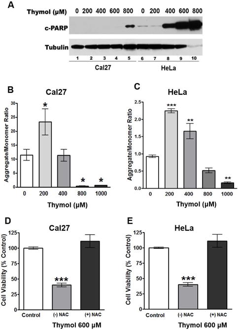
Methods: The antiproliferative effects of thymol in OSCC Cal27 cells were determined by MTS assays. Antitumor effects were evaluated in Cal27- and HeLa-derived mouse xenografts. Calcium imaging, mitochondrial transmembrane potential (ΔΨm) studies, and Western blot analysis of cleaved PARP (c-PARP) evaluated thymol's mechanism-of-action.

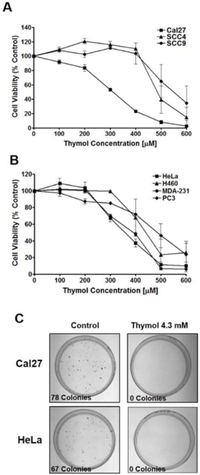
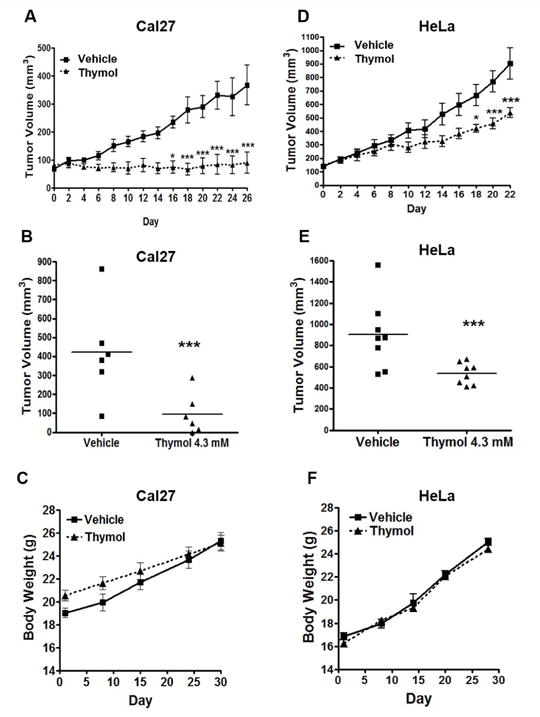
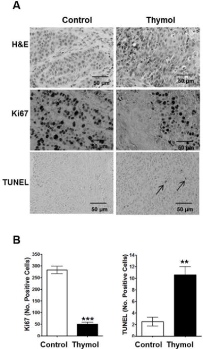
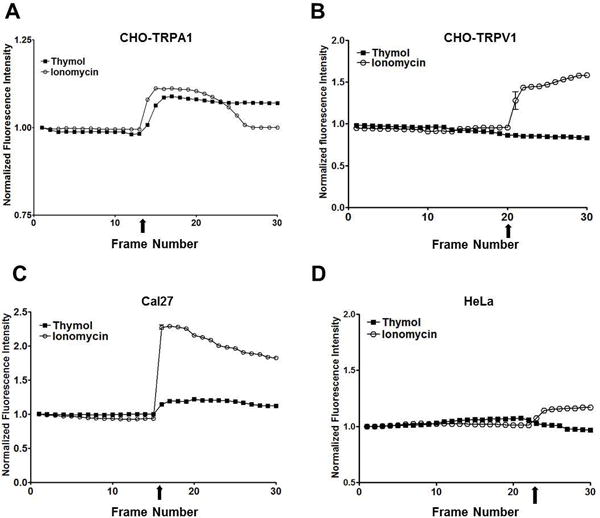
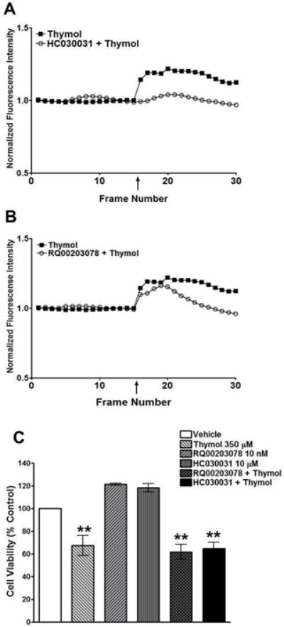
Results: Thymol had significant, long-lasting antiproliferative effects in vitro. In vivo, thymol displayed significant antitumor effects in Cal27-derived tumors. Thymol's anticancer effects were confirmed in HeLa-derived xenografts demonstrating that thymol effects are not tumor-type specific. Calcium imaging verified calcium influx in Cal27 cells that were reversed with the TRPA1 antagonist, HC030031. However, no calcium influx was seen in HeLa cells indicating that TRP channels do not regulate thymol cytotoxicity. This was confirmed using cell viability assays in which pre-treatment with HC030031 had no effect on thymol cytotoxicity. Instead, ΔΨm studies revealed that thymol induces significant ΔΨm depolarization and apoptosis.

Conclusion: Our findings provide the first evidence of thymol's novel antitumor effects against OSCC in vivo, which do not rely on TRPA1 activity. Instead, we show that thymol induces mitochondrial dysfunction and apoptosis and may be efficacious against multiple cancers.

Keywords: TRPA1; apoptosis; mitochondrial dysfunction; oral squamous cell carcinoma; thymol.


Conflict of interest statement

Conflict of Interest Statement

The authors have no conflict of interest.

Figures

Fig. 1
Fig. 1. Thymol inhibits cancer cell proliferation in vitro
A) MTS cell viability assay of OSCC cell lines, Cal27, SCC4, and SCC9 treated with thymol for 48 h revealed GC50 values of 300 μM, 500 μM, and 550 μM respectively (n=4 per group). B) MTS cell viability assay of a panel of cancer cell lines (HeLa, H460, MDA-231, and PC3) treated with thymol for 48 h revealed GC50 values ranging from 350 μM, to 500 μM (n=4 per group). C) Clonogenic assays of Cal27 and HeLa cells 9 days following a 2 min exposure to thymol (4.3 mM; right panels) or vehicle control (left panels).
Fig. 2
Fig. 2. Thymol inhibits Cal27- and HeLa-derived tumor growth in vivo
A & D) Tumor volumes of Cal27 (n=6 per group) and HeLa (n=8 per group) xenografts treated with thymol (4.3 mM) every other day for 26 and 22 days respectively. Significant reduction in Cal27-derived tumor volumes is seen by day 16 (* p<0.05) and in HeLa-derived tumor volumes by day 18 (*p<0.05). B & E) Scatter plot of Cal27 and HeLa tumor volumes respectively. Mean tumor volume for Cal27 control tumors is 423 mm3 whereas mean tumor volume for thymol treated Cal27 tumors is 96 mm3. Mean tumor volume for HeLa control tumors is 903 mm3 whereas mean tumor volume for thymol treated HeLa tumors is 537 mm3. C & F) Changes in body weight over the course of the study is shown for tumor-bearing mice with Cal27-derived xenografts (C) and HeLa-derived xenografts (F).
Fig. 3
Fig. 3. Histopathological analyses of Cal27-derived tumors treated thymol
A) Representative photomicrographs of Cal-27 derived tumors (40 × magnification) stained with H&E or Ki67, and TUNEL assays are shown; apoptotic cells are illustrated by arrows; scale bars = 50 μm. B) Quantification of Ki67 positive cells and apoptotic cells in treated tumors (mean ± SEM; n=3 per group; 3 fields per specimen; **p<0.01, ***p<0.001).
Fig. 4
Fig. 4. Calcium imaging of cell lines treated with thymol (400 μM) or ionomycin positive control (3 μM) show Cal27 cells have TRP channel activity while HeLa cells do not
A) CHO-TRPA1 cells show a thymol-induced calcium influx. B) CHO-TRPV1 cells do not show an influx of calcium in response to thymol. C) Cal27 cells show a thymol-induced calcium influx. D) HeLa cells do not show an influx of calcium in response to thymol.
Fig. 5
Fig. 5. TRPA1 inhibition reduces calcium influx in Cal27 cells whereas the TRPM8 inhibitor does not
A) Calcium imaging of Cal27 cells pre-treated with the TRPA1 antagonist, HC030031 (10 μM), followed by thymol (400 μM) compared to thymol alone (400 μM) show a dramatic reduction in calcium influx. B) Calcium imaging of Cal27 cells pre-treated with the TRPM8 antagonist, RQ00203078 (10 μM), followed by thymol (400 μM) compared to thymol alone (400 μM) have a muted calcium influx even in the presence of TRPM8 inhibition. C) MTS cell viability assay of Cal27 cells treated with thymol (350 μM), HC030031 (10 μM; TRPA1 antagonist), RQ00203078 (10 nM; TRPM8 antagonist), or pre-treated with the TRPA1 or TRPM8 inhibitors followed by thymol (350 μM).
Fig. 6
Fig. 6. Thymol inhibits mitochondrial membrane potential and induces apoptosis
A) Western blot of c-PARP in Cal27 and HeLa cells treated with increasing concentrations of thymol for 48 h show a dose dependent induction of apoptosis. B & C) Cal27 and HeLa cells, respectively, treated with increasing concentrations of thymol for 2 h show significant reduction in mitochondrial membrane potential as measured by JC-1 aggregate to monomer ratio. D & E) MTS cell viability assays of Cal27 and HeLa cells, respectively, following 48 h treatment demonstrate that 10 nM NAC reverses the anti-proliferative effects of thymol.

Similar articles

Cited by

References

    1. Cancer Facts and Figures: Vol.: American Cancer Society. 2015 https://www.cancer.org/research/cancer-facts-statistics/all-cancer-facts... Accessed on 11-13-17.
    1. CONFORTI F, STATTI G, UZUNOV D, et al. Comparative chemical composition and antioxidant activities of wild and cultivated Laurus nobilis L. leaves and Foeniculum vulgare subsp piperitum (Ucria) coutinho seeds. Biol Pharm Bull. 2006;29:2056–64. - PubMed
    1. EL BABILI F, BOUAJILA J, SOUCHARD JP, et al. Oregano: chemical analysis and evaluation of its antimalarial, antioxidant, and cytotoxic activities. J Food Sci. 2011;76:C512–8. - PubMed
    1. MIYAZAWA N, FUJITA A, KUBOTA K. Aroma character impact compounds in Kinokuni mandarin orange (Citrus kinokuni) compared with Satsuma mandarin orange (Citrus unshiu) Biosci Biotechnol Biochem. 2010;74:835–42. - PubMed
    1. OZCAN MM, CHALCHAT JC. Chemical composition and antifungal activity of rosemary (Rosmarinus officinalis L.) oil from Turkey. Int J Food Sci Nutr. 2008;59:691–8. - PubMed

LinkOut - more resources