Review
. 2018 Oct:115:31-42.
doi: 10.1016/j.bone.2018.05.018. Epub 2018 May 16.

Brain to bone: What is the contribution of the brain to skeletal homeostasis?

Affiliations
Review

Brain to bone: What is the contribution of the brain to skeletal homeostasis?

Anna Idelevich et al. Bone. 2018 Oct.

Abstract

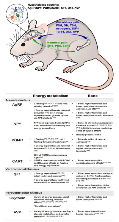
The brain, which governs most, if not all, physiological functions in the body, from the complexities of cognition, learning and memory, to the regulation of basal body temperature, heart rate and breathing, has long been known to affect skeletal health. In particular, the hypothalamus - located at the base of the brain in close proximity to the medial eminence, where the blood-brain-barrier is not as tight as in other regions of the brain but rather "leaky", due to fenestrated capillaries - is exposed to a variety of circulating body cues, such as nutrients (glucose, fatty acids, amino acids), and hormones (insulin, glucagon, leptin, adiponectin) [1-3].Information collected from the body via these peripheral cues is integrated by hypothalamic sensing neurons and glial cells [4-7], which express receptors for these nutrients and hormones, transforming these cues into physiological outputs. Interestingly, many of the same molecules, including leptin, adiponectin and insulin, regulate both energy and skeletal homeostasis. Moreover, they act on a common set of hypothalamic nuclei and their residing neurons, activating endocrine and neuronal systems, which ultimately fine-tune the body to new physiological states. This review will focus exclusively on the brain-to-bone pathway, highlighting the most important anatomical sites within the brain, which are known to affect bone, but not covering the input pathways and molecules informing the brain of the energy and bone metabolic status, covered elsewhere [8-10]. The discussion in each section will present side by side the metabolic and bone-related functions of hypothalamic nuclei, in an attempt to answer some of the long-standing questions of whether energy is affected by bone remodeling and homeostasis and vice versa.

Keywords: Bone; Energy metabolism; Hypothalamus; Pituitary.


Conflict of interest statement

Conflict of interest

The authors declare no conflict of interest

Figures

Figure 1
Figure 1. Arcuate nucleus and ventromedial hypothalamus regulate whole body energy metabolism and bone
Hypothalamus (an area located at the ventral part of the brain, denoted in red) receives peripheral input in the form of body cues – circulating nutrients (glucose, fatty acids, amino acids) and humoral hormones (insulin, glucagon, leptin, adiponectin, ghrelin) which bind to their respective receptors on hypothalamic cells and trigger a metabolic response. This response includes initiation or cessation of appetite, changes in energy expenditure, glucose balance, alterations in fat depots, as well as long-term changes and skeletal homeostasis. Within the hypothalamus, ARC is known to harbor a population of orexigenic AgRP/NPY/GABA expressing neurons co-residing with a population of anorexigenic POMC/CART neurons, although recent studies highlight significant level of heterogeneity within the two. Galanin is also known to be expressed in ARC and is colocolized with AgRP/NPY and POMC/CART neurons. Anterior to ARC lays the VMH marked by the presence of SF-1 neurons. Specific activation of ARC and VMH neurons was shown to trigger endocrine pituitary and/or neuronal (SNS, PNS) downstream response which is ultimately directed at maintaining metabolic and skeletal homeostasis. (Figure constructed using Allen mouse brain volumetric atlas).
Figure 2
Figure 2. Paraventricular nucleus and supraoptic nucleus regulate whole body energy metabolism and bone
Also located in the hypothalamus (an area denoted in red), but anterior to ARC and VMH are PVN and SON areas, which are the principal sites of oxytocin and AVP production. PVN is tightly connected with ARC, as well as other areas in the brain, including the amygdala, brainstem (Raphe nuclei, NTS, VTA.) spinal cord, mesolimbic dopaminergic system (nucleus accumbens) and cortex. Oxytocin and AVP peptides which are produced in the hypothalamus are delivered to the posterior pituitary from which they are discharged into the circulation to control energy balance and skeletal homeostasis. Oxytocin is considered to be anabolic, while AVP is considered to be catabolic to bone. Both hormones are also known to function as neuropeptides binding to their receptors within the brain to affect various aspects of social behavior. (Figure constructed using Allen mouse brain volumetric atlas).
Figure 3
Figure 3. Central output regulating skeletal homeostasis
Hypothalamus utilizes endocrine and neuronal mechanisms to communicate back to the bone. Endocrine route involves all hormones produced by the anterior pituitary gland, including the adrenal, thyroid, gonadal, and somatotropic axis; as well as the posterior pituitary gland, producing oxytocin and AVP. These factors either directly or indirectly, via the release of target tissue hormones, affect both the arms of energy metabolism and bone. – Another route, involves modulation of the activity of the autonomic nervous system, comprising SNS, PNS, (and recently implicated ScNS), which activate adrenergic and/or cholinergic receptors located on bone cells, changing bone formation and resorption to maintain skeletal homeostasis.

References

    1. Haddad-Tóvolli R, Dragano NRV, Ramalho AFS, Velloso LA. Development and Function of the Blood-Brain Barrier in the Context of Metabolic Control. Front Neurosci. 2017;11:224. - PMC - PubMed
    1. Magnan C, Levin BE, Luquet S. Brain lipid sensing and the neural control of energy balance. Mol Cell Endocrinol. 2015 Dec;418:3–8. - PubMed
    1. Lam TKT, Schwartz GJ, Rossetti L. Hypothalamic sensing of fatty acids. Nat Neurosci. 2005 May;8(5):579–84. - PubMed
    1. Leloup C, Allard C, Carneiro L, Fioramonti X, Collins S, Pénicaud L. Glucose and hypothalamic astrocytes: More than a fueling role? Neuroscience. 2015 Jun;323:110–120. - PubMed
    1. Steinbusch L, Labouèbe G, Thorens B. Brain glucose sensing in homeostatic and hedonic regulation. Trends Endocrinol Metab. 2015 Sep;26(9):455–466. - PubMed

Publication types

LinkOut - more resources