

Altered mitochondrial acetylation profiles in a kainic acid model of temporal lobe epilepsy
- PMID: 29778462
- PMCID: PMC6082368
- DOI: 10.1016/j.freeradbiomed.2018.05.063
Altered mitochondrial acetylation profiles in a kainic acid model of temporal lobe epilepsy
Abstract
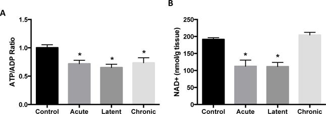
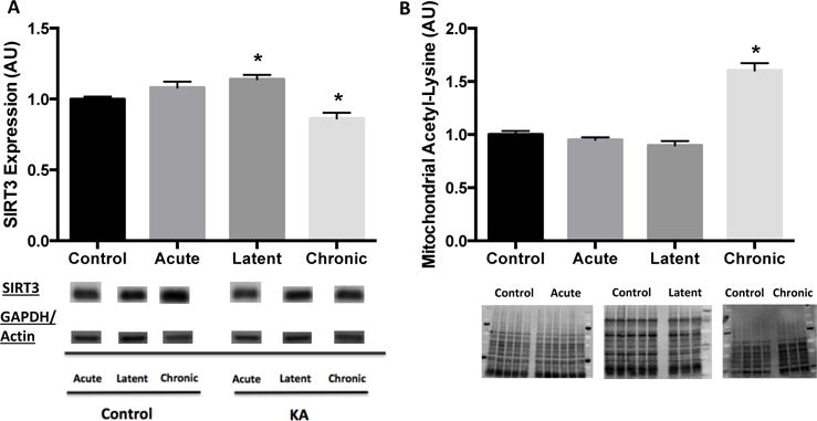
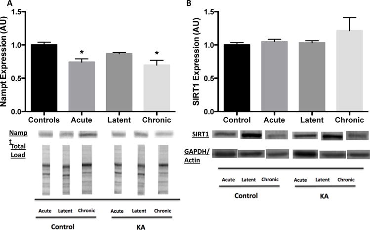
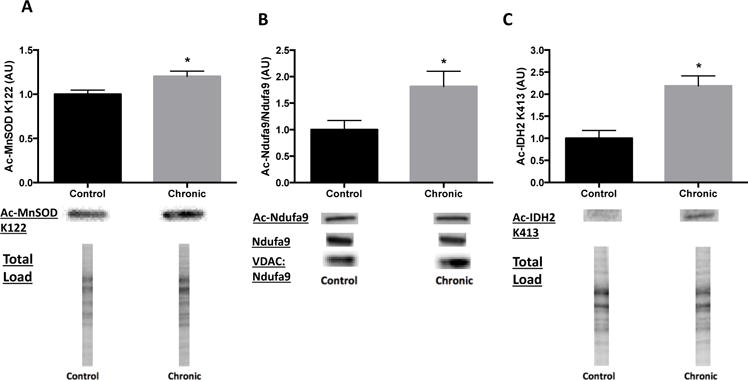
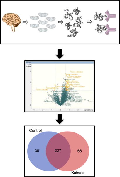
Impaired bioenergetics and oxidative damage in the mitochondria are implicated in the etiology of temporal lobe epilepsy, and hyperacetylation of mitochondrial proteins has recently emerged as a critical negative regulator of mitochondrial functions. However, the roles of mitochondrial acetylation and activity of the primary mitochondrial deacetylase, SIRT3, have not been explored in acquired epilepsy. We investigated changes in mitochondrial acetylation and SIRT3 activity in the development of chronic epilepsy in the kainic acid rat model of TLE. Hippocampal measurements were made at 48 h, 1 week and 12 weeks corresponding to the acute, latent and chronic stages of epileptogenesis. Assessment of hippocampal bioenergetics demonstrated a ≥ 27% decrease in the ATP/ADP ratio at all phases of epileptogenesis (p < 0.05), whereas cellular NAD+ levels were decreased by ≥ 41% in the acute and latent time points (p < 0.05), but not in chronically epileptic rats. In spontaneously epileptic rats, we found decreased protein expression of SIRT3 and a 60% increase in global mitochondrial acetylation, as well as enhanced acetylation of the known SIRT3 substrates MnSOD, Ndufa9 of Complex I and IDH2 (all p < 0.05), suggesting SIRT3 dysfunction in chronic epilepsy. Mass spectrometry-based acetylomics investigation of hippocampal mitochondria demonstrated a 79% increase in unique acetylated proteins from rats in the chronic phase vs. controls. Pathway analysis identified numerous mitochondrial bioenergetic pathways affected by mitochondrial acetylation. These results suggest SIRT3 dysfunction and aberrant protein acetylation may contribute to mitochondrial dysfunction in chronic epilepsy.
Keywords: Acetylation; Epilepsy; Mass spectrometry; Mitochondria; Proteomics; SIRT3.
Copyright © 2018 Elsevier Inc. All rights reserved.
Figures
Similar articles
-
Mitochondrial respiration deficits driven by reactive oxygen species in experimental temporal lobe epilepsy.Neurobiol Dis. 2015 Mar;75:151-8. doi: 10.1016/j.nbd.2014.12.025. Epub 2015 Jan 17. Neurobiol Dis. 2015. PMID: 25600213 Free PMC article.
-
Wnt/β-catenin signalling pathway mediated aberrant hippocampal neurogenesis in kainic acid-induced epilepsy.Cell Biochem Funct. 2017 Oct;35(7):472-476. doi: 10.1002/cbf.3306. Epub 2017 Oct 19. Cell Biochem Funct. 2017. PMID: 29052243
-
Altered cardiac structure and function is related to seizure frequency in a rat model of chronic acquired temporal lobe epilepsy.Neurobiol Dis. 2021 Nov;159:105505. doi: 10.1016/j.nbd.2021.105505. Epub 2021 Sep 11. Neurobiol Dis. 2021. PMID: 34520843
-
The kainic acid model of temporal lobe epilepsy.Neurosci Biobehav Rev. 2013 Dec;37(10 Pt 2):2887-99. doi: 10.1016/j.neubiorev.2013.10.011. Epub 2013 Oct 30. Neurosci Biobehav Rev. 2013. PMID: 24184743 Free PMC article. Review.
-
Mitochondrial involvement and oxidative stress in temporal lobe epilepsy.Free Radic Biol Med. 2013 Sep;62:121-131. doi: 10.1016/j.freeradbiomed.2013.02.002. Epub 2013 Feb 11. Free Radic Biol Med. 2013. PMID: 23411150 Free PMC article. Review.
Cited by
-
The Brain Protein Acylation System Responds to Seizures in the Rat Model of PTZ-Induced Epilepsy.Int J Mol Sci. 2022 Oct 14;23(20):12302. doi: 10.3390/ijms232012302. Int J Mol Sci. 2022. PMID: 36293175 Free PMC article.
-
A Compared Study of Eicosapentaenoic Acid and Docosahexaenoic Acid in Improving Seizure-Induced Cognitive Deficiency in a Pentylenetetrazol-Kindling Young Mice Model.Mar Drugs. 2023 Aug 24;21(9):464. doi: 10.3390/md21090464. Mar Drugs. 2023. PMID: 37755077 Free PMC article.
-
Antioxidants Targeting Mitochondrial Oxidative Stress: Promising Neuroprotectants for Epilepsy.Oxid Med Cell Longev. 2020 Nov 25;2020:6687185. doi: 10.1155/2020/6687185. eCollection 2020. Oxid Med Cell Longev. 2020. PMID: 33299529 Free PMC article. Review.
-
Neuronal SIRT3 Deletion Predisposes to Female-Specific Alterations in Cellular Metabolism, Memory, and Network Excitability.J Neurosci. 2023 Mar 8;43(10):1845-1857. doi: 10.1523/JNEUROSCI.1259-22.2023. Epub 2023 Feb 9. J Neurosci. 2023. PMID: 36759193 Free PMC article.
-
Mitochondrial dysfunction and NLRP3 inflammasome activation in drug-resistant epilepsy: emerging insights and mitochondrial-targeted therapeutic strategies.Naunyn Schmiedebergs Arch Pharmacol. 2025 Jul 23. doi: 10.1007/s00210-025-04468-2. Online ahead of print. Naunyn Schmiedebergs Arch Pharmacol. 2025. PMID: 40699241 Review.
References
-
- Botker HE, Kimose HH, Helligso P, Nielsen TT. Analytical evaluation of high energy phosphate determination by high performance liquid chromatography in myocardial tissue. Journal of molecular and cellular cardiology. 1994;26:41–48. - PubMed
Publication types
MeSH terms
Substances
Grants and funding
LinkOut - more resources
Full Text Sources
Other Literature Sources
Miscellaneous
