

Limbic system structure volumes and associated neurocognitive functioning in former NFL players
- PMID: 29779184
- PMCID: PMC6854905
- DOI: 10.1007/s11682-018-9895-z
Limbic system structure volumes and associated neurocognitive functioning in former NFL players
Abstract
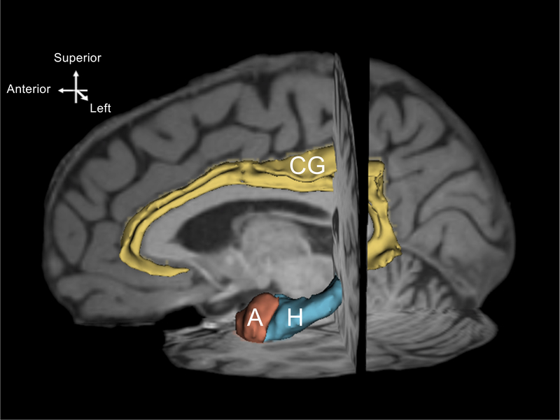
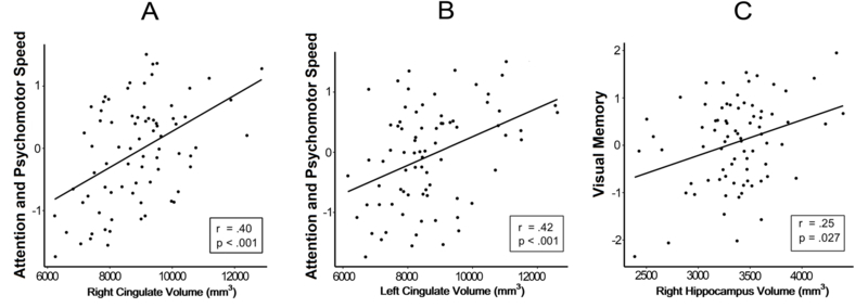
Chronic traumatic encephalopathy (CTE) is a neurodegenerative disease associated with exposure to repetitive head impacts. CTE has been linked to disruptions in cognition, mood, and behavior. Unfortunately, the diagnosis of CTE can only be made post-mortem. Neuropathological evidence suggests limbic structures may provide an opportunity to characterize CTE in the living. Using 3 T magnetic resonance imaging, we compared select limbic brain regional volumes - the amygdala, hippocampus, and cingulate gyrus - between symptomatic former National Football League (NFL) players (n = 86) and controls (n = 22). Moreover, within the group of former NFL players, we examined the relationship between those limbic structures and neurobehavioral functioning (n = 75). The former NFL group comprised eighty-six men (mean age = 55.2 ± 8.0 years) with at least 12 years of organized football experience, at least 2 years of active participation in the NFL, and self-reported declines in cognition, mood, and behavior within the last 6 months. The control group consisted of men (mean age = 57.0 ± 6.6 years) with no history of contact-sport involvement or traumatic brain injury. All control participants provided neurobehavioral data. Compared to controls, former NFL players exhibited reduced volumes of the amygdala, hippocampus, and cingulate gyrus. Within the NFL group, reduced bilateral cingulate gyrus volume was associated with worse attention and psychomotor speed (r = 0.4 (right), r = 0.42 (left); both p < 0.001), while decreased right hippocampal volume was associated with worse visual memory (r = 0.25, p = 0.027). Reduced volumes of limbic system structures in former NFL players are associated with neurocognitive features of CTE. Volume reductions in the amygdala, hippocampus, and cingulate gyrus may be potential biomarkers of neurodegeneration in those at risk for CTE.
Keywords: Chronic Traumatic Encephalopathy; Hippocampus; Limbic System; Repetitive Head Impacts; Volumetric MRI.
Conflict of interest statement
Figures
References
-
- Amaral DG, Capitanio JP, Jourdain M, Mason WA, Mendoza SP, & Prather M. (2003). The amygdala: is it an essential component of the neural network for social cognition? Neuropsychologia, 41(2), 235–240. - PubMed
-
- Bernick C, Banks SJ, Shin W, Obuchowski N, Butler S, Noback M, et al. (2015). Repeated head trauma is associated with smaller thalamic volumes and slower processing speed: the Professional Fighters’ Brain Health Study. Br J Sports Med, 49(15), 1007–1011, doi:10.1136/bjsports-2014-093877. - DOI - PMC - PubMed
MeSH terms
Grants and funding
- T32GM074905/NH/NIH HHS/United States
- 1F32NS096803-01/NH/NIH HHS/United States
- T32 GM074905/GM/NIGMS NIH HHS/United States
- R01 NS 078337/NH/NIH HHS/United States
- U01 NS093334/NS/NINDS NIH HHS/United States
- K23 NS102399/NS/NINDS NIH HHS/United States
- UL1-TR000157/NH/NIH HHS/United States
- UL1 TR000157/TR/NCATS NIH HHS/United States
- P30 AG019610/AG/NIA NIH HHS/United States
- F31 NS081957/NS/NINDS NIH HHS/United States
- F32 NS096803/NS/NINDS NIH HHS/United States
- P30 AG013846/AG/NIA NIH HHS/United States
- R01 NS100952/NS/NINDS NIH HHS/United States
- P30 AG13846/NH/NIH HHS/United States
- F31 NS 081957/NH/NIH HHS/United States
- R01 NS078337/NS/NINDS NIH HHS/United States
- P41 EB015902/NH/NIH HHS/United States
- P41 EB015902/EB/NIBIB NIH HHS/United States
LinkOut - more resources
Full Text Sources
Other Literature Sources
