Review
. 2018 Sep;125(1-2):18-37.
doi: 10.1016/j.ymgme.2018.05.004. Epub 2018 May 15.

Molecular genetics and metabolism, special edition: Diagnosis, diagnosis and prognosis of Mucopolysaccharidosis IVA

Affiliations
Review

Molecular genetics and metabolism, special edition: Diagnosis, diagnosis and prognosis of Mucopolysaccharidosis IVA

Hira Peracha et al. Mol Genet Metab. 2018 Sep.

Abstract

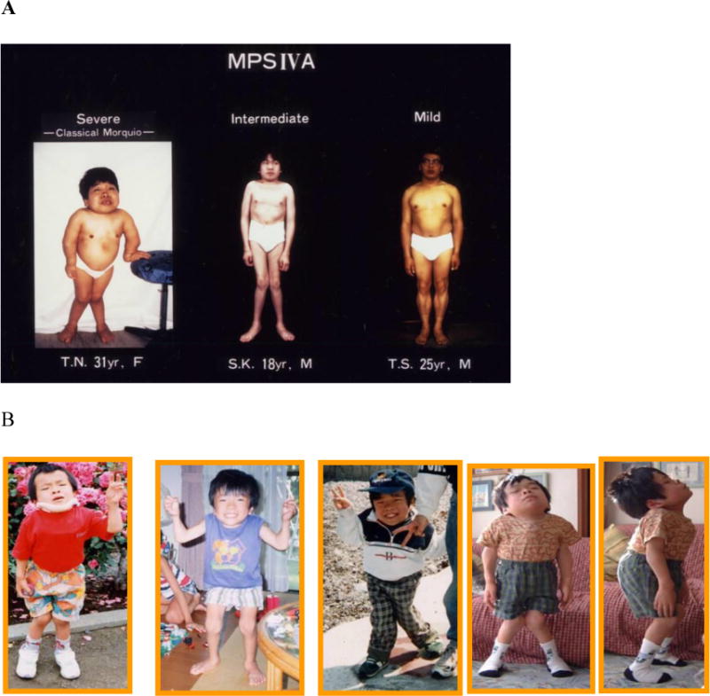
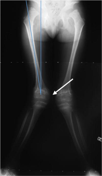
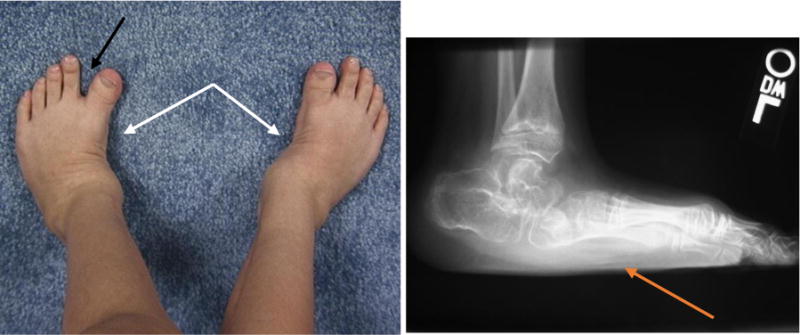
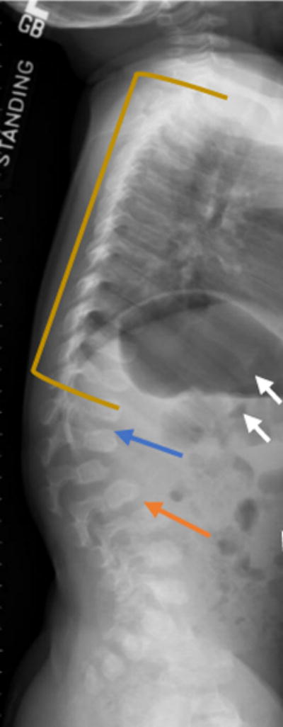
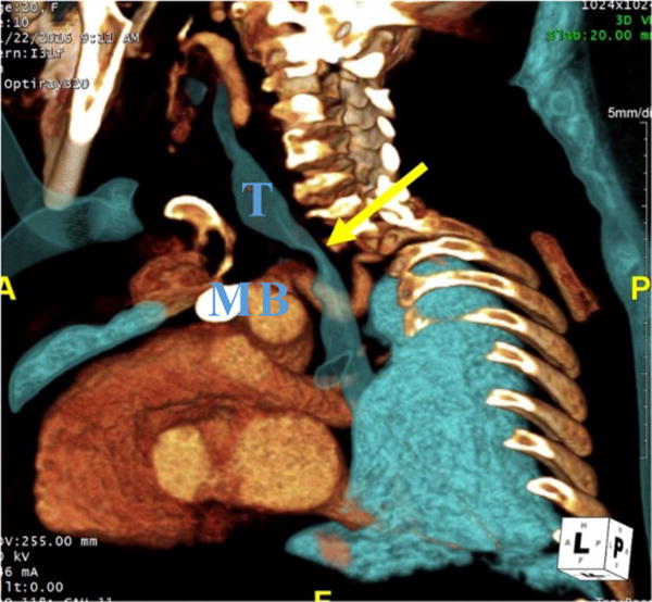
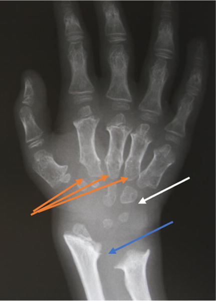
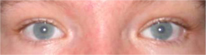
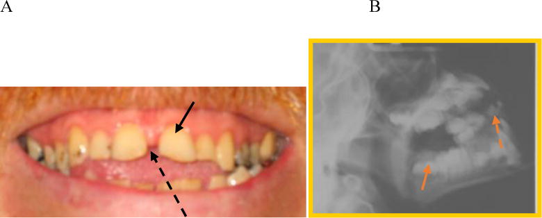
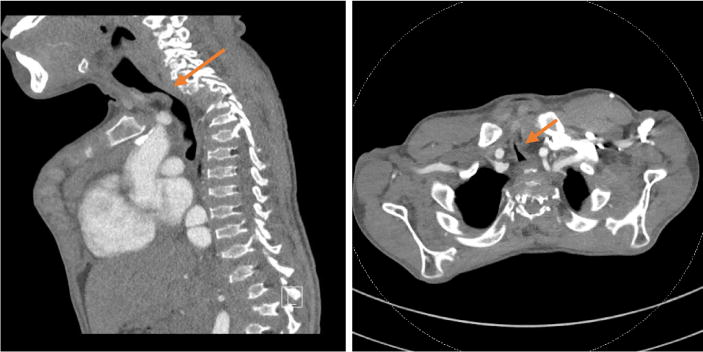
Mucopolysaccharidosis IVA (MPS IVA, Morquio A syndrome) is an autosomal recessive disorder caused by the deficiency of N-acetylgalactosamine-6-sulfate sulfatase. Deficiency of this enzyme leads to the accumulation of specific glycosaminoglycans (GAGs), chondroitin-6-sulfate (C6S) and keratan sulfate (KS), which are mainly synthesized in the cartilage. Therefore, the substrates are stored primarily in the cartilage and its extracellular matrix (ECM), leading to a direct impact on bone development and successive systemic skeletal spondylepiphyseal dysplasia. The skeletal-related symptoms for MPS IVA include short stature with short neck and trunk, odontoid hypoplasia, spinal cord compression, tracheal obstruction, obstructive airway, pectus carinatum, restrictive lung, kyphoscoliosis, platyspondyly, coxa valga, genu valgum, waddling gait, and laxity of joints. The degree of imbalance of growth in bone and other organs and tissues largely contributes to unique skeletal dysplasia and clinical severity. Diagnosis of MPS IVA needs clinical, radiographic, and laboratory testing to make a complete conclusion. To diagnose MPS IVA, total urinary GAG analysis which has been used is problematic since the values overlap with those in age-matched controls. Currently, urinary and blood KS and C6S, the enzyme activity of GALNS, and GALNS molecular analysis are used for diagnosis and prognosis of clinical phenotype in MPS IVA. MPS IVA can be diagnosed with unique characters although this disorder relates closely to other disorders in some characteristics. In this review article, we comprehensively describe clinical, radiographic, biochemical, and molecular diagnosis and clinical assessment tests for MPS IVA. We also compare MPS IVA to other closely related disorders to differentiate MPS IVA. Overall, imbalance of growth in MPS IVA patients underlies unique skeletal manifestations leading to a critical indicator for diagnosis.

Keywords: Chondroitin-6-sulfate; Keratan sulfate; Mucopolysaccharidosis IVA; N-acetylgalactosamine-6-sulfate; Spondyloepiphyseal dysplasia.


Conflict of interest statement

Conflict of Interest:

All the authors contributed to the Review Article and had no conflict of interest with any other party. Hira Peracha, Kazuki Sawamoto, Lauren Averill, Heidi Kecskemethy, Mary Theroux, Mihir Thacker, Kyoko Nagao, Christian Pizarro, William Mackenzie, Hironori Kobayashi, Seiji Yamaguchi, Yasuyuki Suzuki, Kenji Orii, Tadao Orii, Toshiyuki Fukao, Shunji Tomatsu declare that they have no conflict of interests.

Figures

Figure 1
Figure 1
Patients with mild to severe form of MPS IVA. A. Images of patients with distinct differences in clinical features from mild form, immediate form, and severe form of MPS IVA (adapted from Educational CD for Morquio and permitted by Carol Ann Foundation). B. Images of a patient with severe form of MPS IVA. From left to right: patient age is 7, 8, 11, 16, and 16 years (adapted from Educational CD for Morquio and permitted by Carol Ann Foundation) [33].
Figure 2
Figure 2
Time course of clinical symptoms in MPS IVA
Figure 3
Figure 3
Lateral radiographs of the cervical spine in flexion (a) and extension (B) show atlantoaxial instablity in an 8-year-old female patient with MPS IVA. The anterior arch of C1 moves anteriorly in flexion (arrow) in relation to C2. There is hypoplasia of the dens (dashed arrow) and generalized platyspondyly.
Figure 4
Figure 4
Erect frontal radiograph of the hips from a patient with MPS IVA. Shown here is progressive proximal and lateral migration (blue arrow) as well as the flattening and eventual disappearance of the proximal femoral epiphysis (white arrows).
Figure 5
Figure 5
Erect frontal radiographs of the lower extremities of a patient with genu valgum.
Figure 6
Figure 6
Feet of a patient with MPS IVA. Photograph of the feet (a) and lateral radiograph of the left foot (b) of this MPS IVA patient show skew foot posture in both feet (white arrows), pronated pes planus (orange arrow), and increased sandal gap between the first and second toes (black arrow).
Figure 7
Figure 7
Lateral radiograph of the spine of a two-year-old MPS IVA patient with vertebral wedging (blue arrow). Platyspondyly (yellow line), anterior inferior beaking (orange arrow), rib flaring (white arrows) are seen in this X-ray radiograph of a patient with MPS IVA.
Figure 8
Figure 8
CT angiography (CTA) of a 20-year-old woman with MPS IVA. 3D oblique reconstruction of CTA demonstrates marked narrowing of the trachea at the thoracic inlet (yellow arrow). T– Trachea, B- Brachiocephalic artery, M- Manubrium.
Figure 9
Figure 9
X-ray radiograph of an MPS IVA patient hand. Clearly seen are the tapering of the proximal portion for the metacarpals (orange arrows), small irregular carpal bones (white arrow), and the distal portion of the radius being tilted toward the ulna (blue arrow).
Figure 10
Figure 10
Eyes of a patient with MPS IVA. Image of a 24-year-old patient with MPS IVA shows fine stromal corneal clouding (adapted from Educational CD for Morquio and permitted by Carol Ann Foundation).
Figure 11
Figure 11
Teeth of a patient with MPS IVA. A. Image of a 24-year-old patient with widely spaced teeth (black arrow) and spade shaped incisors (black dotted arrow) (adapted from Educational CD for Morquio and permitted by Carol Ann Foundation). B. X-ray of a MPS IVA patient with concave occlulsal sufraces (orange arrow) and spade-shaped incisors (orange dotted arrow).
Figure 12
Figure 12
Sagittal (a) and axial (b) CT images of a 16-year-old boy with MPS IVA focal severe tracheal narrowing at the thoracic inlet (arrows). Tracheal cross-sectional area measures 10 mm2 at the thoracic inlet and 110 mm2 intrathoracic below the area of narrowing.
Figure 13
Figure 13
DXA image of the spine of a MPS IVA patient. Lumbar spine DXA requires careful and precise measurement of vertebral body levels and margins (Adapted from Kecskemethy et al. Mol Genet Metab; 2017, 144-149) [145].

References

    1. Gentili C, Cancedda R. Cartilage and bone extracellular matrix. Curr Pharm Des. 2009;15:1334–48. http://www.ncbi.nlm.nih.gov/pubmed/19355972 (accessed October 28, 2017) - PubMed
    1. Alliston T. Chondroitin sulfate and growth factor signaling in the skeleton: Possible links to MPS VI. J Pediatr Rehabil Med. 2010;3:129–38. doi: 10.3233/PRM-2010-0117. - DOI - PMC - PubMed
    1. Khan S, Alméciga-Díaz CJ, Sawamoto K, Mackenzie WG, Theroux MC, Pizarro C, Mason RW, Orii T, Tomatsu S. Mucopolysaccharidosis IVA and glycosaminoglycans. Mol Genet Metab. 2017;120:78–95. doi: 10.1016/j.ymgme.2016.11.007. - DOI - PMC - PubMed
    1. Staprans I. Isolation and characterization of glycosaminoglycans in human plasma. J Clin Invest. 1985;76:1984–91. doi: 10.1172/JCI112198. - DOI - PMC - PubMed
    1. Morquio L. Sur une forme de dystrophie osseuse familiale. Bull Soc Pediatr Paris. 1929;27:145–52.

Publication types

LinkOut - more resources