. 2018 Sep-Oct;11(5):1140-1150.
doi: 10.1016/j.brs.2018.05.008. Epub 2018 May 12.

Action potential initiation, propagation, and cortical invasion in the hyperdirect pathway during subthalamic deep brain stimulation

Affiliations

Action potential initiation, propagation, and cortical invasion in the hyperdirect pathway during subthalamic deep brain stimulation

Ross W Anderson et al. Brain Stimul. 2018 Sep-Oct.

Abstract

Background: High frequency (∼130 Hz) deep brain stimulation (DBS) of the subthalamic region is an established clinical therapy for the treatment of late stage Parkinson's disease (PD). Direct modulation of the hyperdirect pathway, defined as cortical layer V pyramidal neurons that send an axon collateral to the subthalamic nucleus (STN), has emerged as a possible component of the therapeutic mechanisms. However, numerous questions remain to be addressed on the basic biophysics of hyperdirect pathway stimulation.

Objective: Quantify action potential (AP) initiation, propagation, and cortical invasion in hyperdirect neurons during subthalamic stimulation.

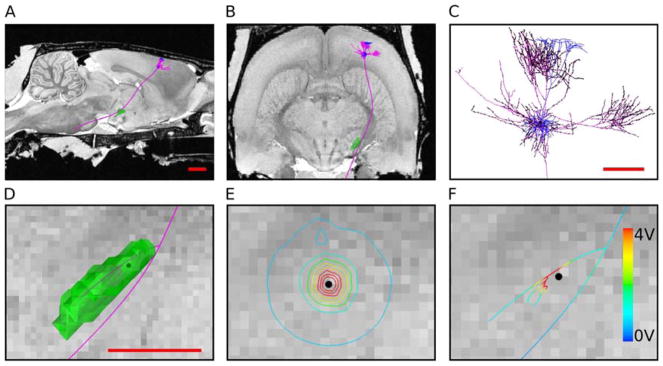
Methods: We developed an anatomically and electrically detailed computational model of hyperdirect neuron stimulation with explicit representation of the stimulating electric field, axonal response, AP propagation, and synaptic transmission.

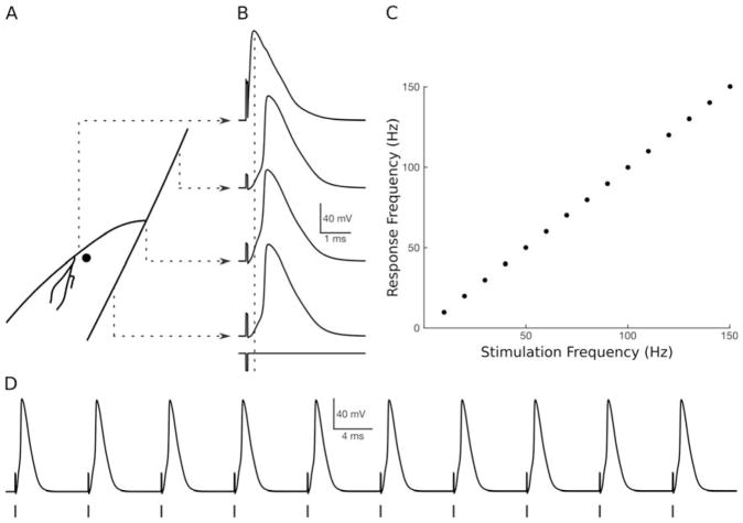
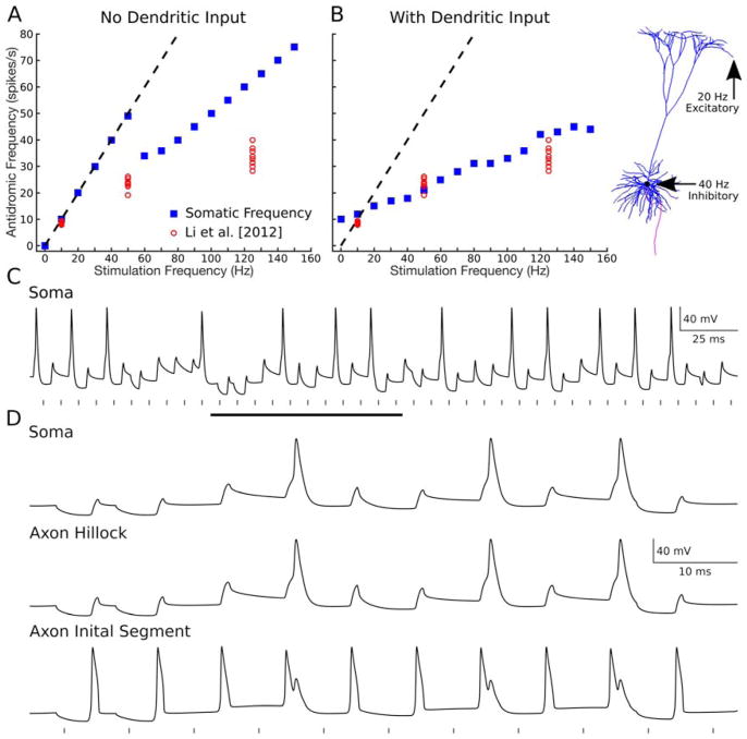
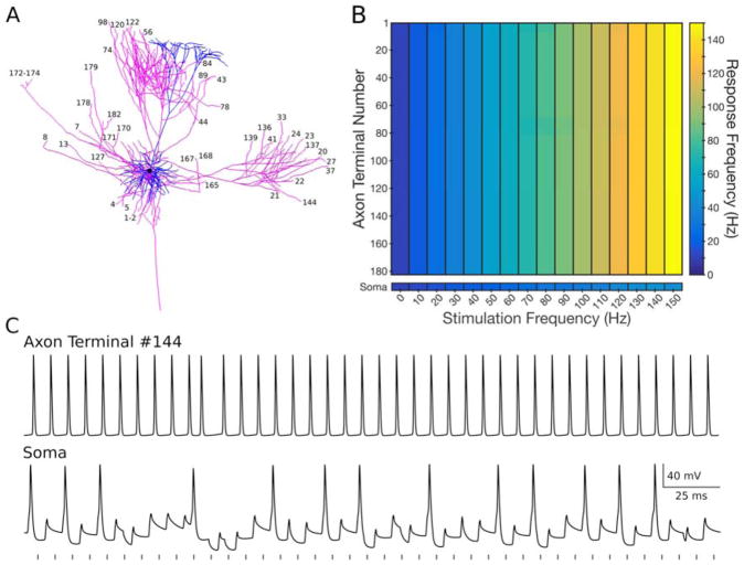
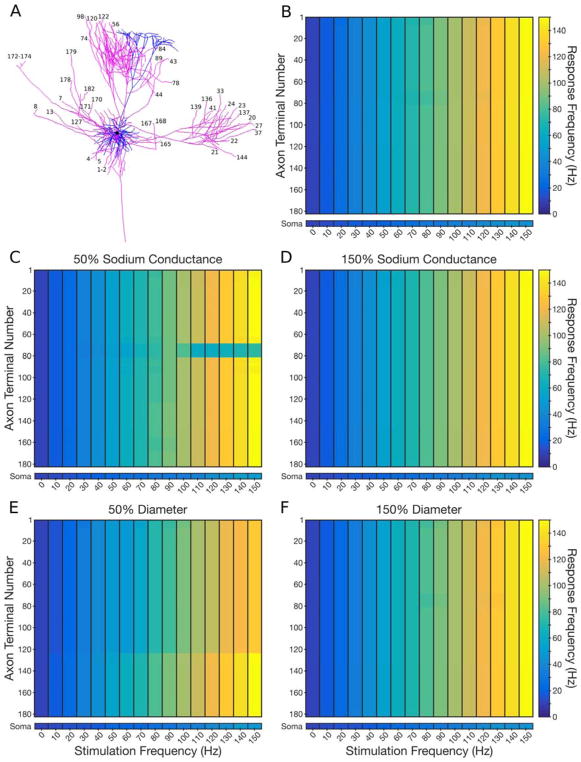
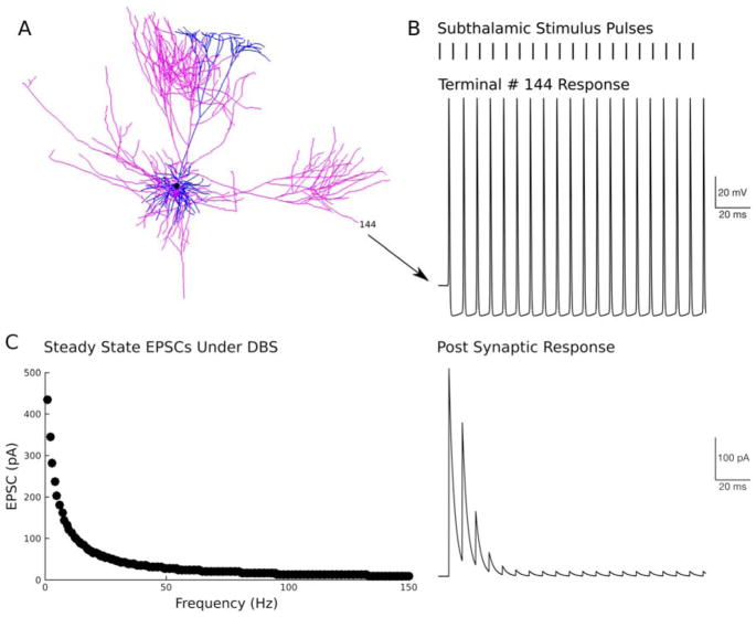
Results: We found robust AP propagation throughout the complex axonal arbor of the hyperdirect neuron. Even at therapeutic DBS frequencies, stimulation induced APs could reach all of the intracortical axon terminals with ∼100% fidelity. The functional result of this high frequency axonal driving of the thousands of synaptic connections made by each directly stimulated hyperdirect neuron is a profound synaptic suppression that would effectively disconnect the neuron from the cortical circuitry.

Conclusions: The synaptic suppression hypothesis integrates the fundamental biophysics of electrical stimulation, axonal transmission, and synaptic physiology to explain a generic mechanism of DBS.

Keywords: Corticofugal axon; Pyramidal neuron; Subthalamic nucleus.


Conflict of interest statement

Conflict of Interest Statement: CCM is a paid consultant for Boston Scientific Neuromodulation and Kernel, as well as a shareholder in the following companies: Surgical Information Sciences, Inc.; Autonomic Technologies, Inc.; Cardionomic, Inc.; Enspire DBS, Inc.; Neuros Medical, Inc.

Figures

Figure 1
Figure 1
Subthalamic stimulation of a hyperdirect neuron. A) Sagittal and B) coronal view of the model system. The layer V pyramidal neuron (blue) is positioned in motor cortex and connected to the intracortical axonal arbor (pink). A corticofugal axon projects down internal capsule and provides a branching collateral to the subthalamic nucleus (STN) (green). C) Close-up view of the cortical region. Black dots represent synaptic bouton locations. D–F) Close-up view of the subthalamic region. Black dot represents the electrode location. Each panel shows the same field of view. D) Anatomical image of the STN and hyperdirect collateral. E) Electrical image of the voltage distribution generated by the stimulus. F) Extracellular voltage distribution applied to the neuron model. All scale bars = 500 μm.
Figure 2
Figure 2
Action potential initiation. A) Anatomical schematic of the electrode location relative to the hyperdirect axon. B) Transmembrane voltage responses to a suprathreshold stimulus pulse. Resting membrane potential of −70 mV. C) Action potential response frequency as a function of the stimulation frequency. D) Transmembrane voltage response at the corticofugal branch point to the 130 Hz stimulus train.
Figure 3
Figure 3
Cortical invasion. A) Action potential propagation along corticofugal axon. Colors denote the transmembrane voltage at the specified time points following the stimulus pulse. B) Antidromic invasion of the cortical model components. C) Times that action potentials reach the terminal ends of the intracortical axonal arbor for a single pulse or trains of pulses.
Figure 4
Figure 4
Somatic firing. A, B) Antidromic action potential invasion of the soma as a function of the subthalamic stimulation frequency. A) Model results with no synaptic inputs to the dendrites. B) Model results with simulated excitatory and inhibitory synaptic inputs to the dendrites. In vivo experimental results of Li et al. [2012] plotted for reference. C) Transmembrane voltage of the soma from B with a stimulation frequency of 130 Hz. D) Transmembrane voltage of the soma, axon hillock, and initial segment for the time window marked with the black bar in C.
Figure 5
Figure 5
Intracortical axon firing. A) Numbering of the various axon terminations in cortex. B) Response frequency of the axon terminations and the soma as a function of the subthalamic stimulation frequency. C) Example transmembrane voltage traces at 130 Hz.
Figure 6
Figure 6
Sensitivity analysis. A) Numbering of the various axon terminals. B) Response frequency of the axon terminals and the soma as a function of the subthalamic stimulation frequency for the default model. C) Response frequency after decreasing the sodium channel conductance of the intracortical axonal arbor by 50%. D) Response frequency after increasing the sodium channel conductance of the intracortical axonal arbor by 50%. E) Response frequency after decreasing the diameter of the intracortical axonal arbor by 50%. F) Response frequency after increasing the diameter of the intracortical axonal arbor by 50%.
Figure 7
Figure 7
Synaptic suppression. A) Neuron model. B) Subthalamic stimuli at 130 Hz generate action potentials that faithfully invade terminal #144. The simulated excitatory post-synaptic current (EPSC) generated by each pre-synaptic signal rapidly suppresses in response to the 130 Hz driving. C) The steady-state EPSC as a function of the stimulation frequency.

Similar articles

Cited by

References

    1. Li Q, Qian ZM, Arbuthnott GW, Ke Y, Yung WH. Cortical effects of deep brain stimulation: implications for pathogenesis and treatment of Parkinson disease. JAMA Neurol. 2014;71(1):100–3. - PubMed
    1. Nambu A, Tokuno H, Inase M, Takada M. Corticosubthalamic input zones from forelimb representations of the dorsal and ventral divisions of the premotor cortex in the macaque monkey: comparison with the input zones from the primary motor cortex and the supplementary motor area. Neurosci Lett. 1997;239(1):13–6. - PubMed
    1. Kita T, Kita H. The subthalamic nucleus is one of multiple innervation sites for long-range corticofugal axons: a single-axon tracing study in the rat. J Neurosci. 2012;32(17):5990–9. - PMC - PubMed
    1. Haynes WI, Haber SN. The organization of prefrontal-subthalamic inputs in primates provides an anatomical substrate for both functional specificity and integration: implications for Basal Ganglia models and deep brain stimulation. J Neurosci. 2013;33(11):4804–14. - PMC - PubMed
    1. Kita T, Osten P, Kita H. Rat subthalamic nucleus and zona incerta share extensively overlapped representations of cortical functional territories. J Comp Neurol. 2014;522(18):4043–56. - PMC - PubMed

Publication types