. 2018 Jun;5(6):e241-e251.
doi: 10.1016/S2352-3026(18)30053-X. Epub 2018 May 17.

Automated typing of red blood cell and platelet antigens: a whole-genome sequencing study

Collaborators, Affiliations

Automated typing of red blood cell and platelet antigens: a whole-genome sequencing study

William J Lane et al. Lancet Haematol. 2018 Jun.

Abstract

Background: There are more than 300 known red blood cell (RBC) antigens and 33 platelet antigens that differ between individuals. Sensitisation to antigens is a serious complication that can occur in prenatal medicine and after blood transfusion, particularly for patients who require multiple transfusions. Although pre-transfusion compatibility testing largely relies on serological methods, reagents are not available for many antigens. Methods based on single-nucleotide polymorphism (SNP) arrays have been used, but typing for ABO and Rh-the most important blood groups-cannot be done with SNP typing alone. We aimed to develop a novel method based on whole-genome sequencing to identify RBC and platelet antigens.

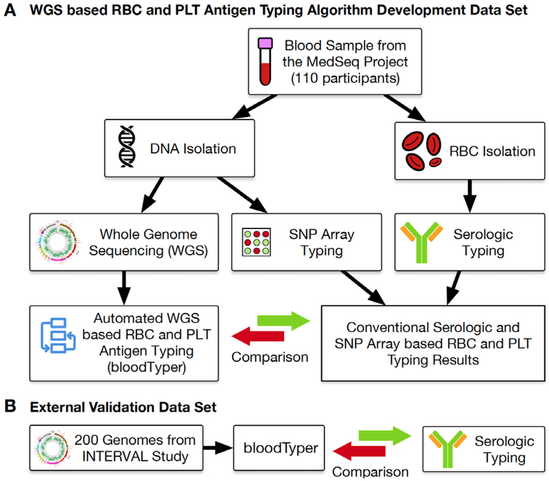
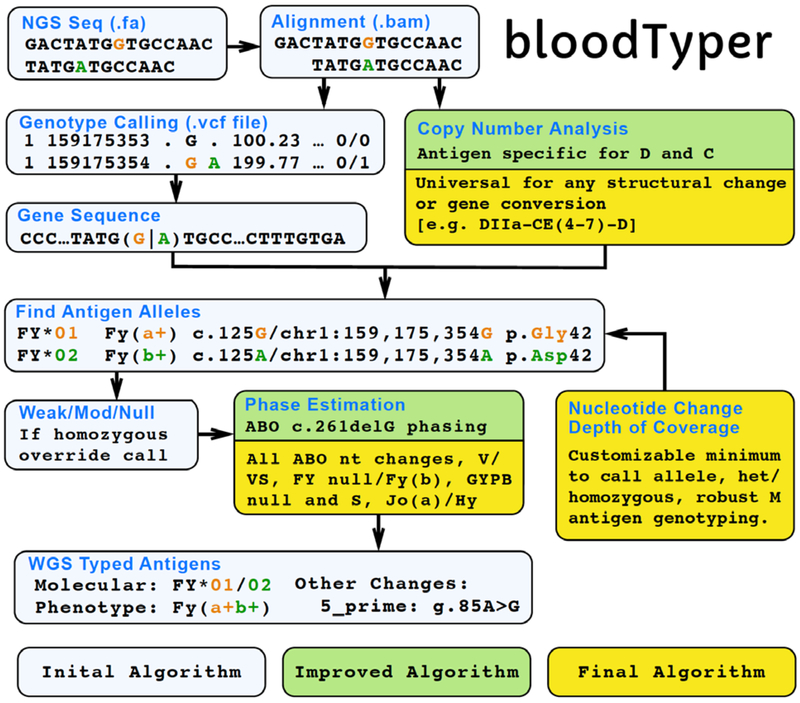
Methods: This whole-genome sequencing study is a subanalysis of data from patients in the whole-genome sequencing arm of the MedSeq Project randomised controlled trial (NCT01736566) with no measured patient outcomes. We created a database of molecular changes in RBC and platelet antigens and developed an automated antigen-typing algorithm based on whole-genome sequencing (bloodTyper). This algorithm was iteratively improved to address cis-trans haplotype ambiguities and homologous gene alignments. Whole-genome sequencing data from 110 MedSeq participants (30 × depth) were used to initially validate bloodTyper through comparison with conventional serology and SNP methods for typing of 38 RBC antigens in 12 blood-group systems and 22 human platelet antigens. bloodTyper was further validated with whole-genome sequencing data from 200 INTERVAL trial participants (15 × depth) with serological comparisons.

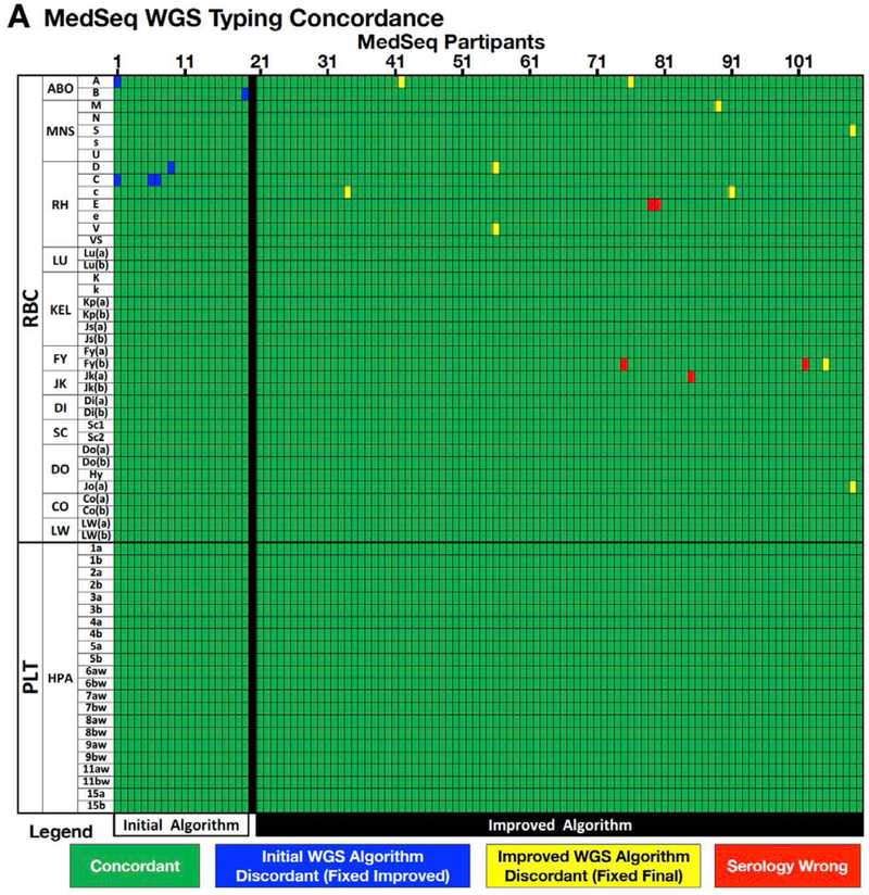
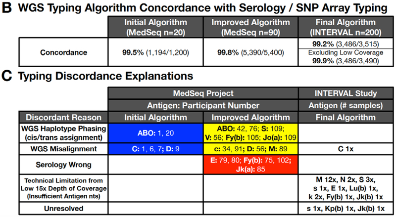
Findings: We iteratively improved bloodTyper by comparing its typing results with conventional serological and SNP typing in three rounds of testing. The initial whole-genome sequencing typing algorithm was 99·5% concordant across the first 20 MedSeq genomes. Addressing discordances led to development of an improved algorithm that was 99·8% concordant for the remaining 90 MedSeq genomes. Additional modifications led to the final algorithm, which was 99·2% concordant across 200 INTERVAL genomes (or 99·9% after adjustment for the lower depth of coverage).

Interpretation: By enabling more precise antigen-matching of patients with blood donors, antigen typing based on whole-genome sequencing provides a novel approach to improve transfusion outcomes with the potential to transform the practice of transfusion medicine.

Funding: National Human Genome Research Institute, Doris Duke Charitable Foundation, National Health Service Blood and Transplant, National Institute for Health Research, and Wellcome Trust.


Figures

Figure 1.
Figure 1.
WGS Study Overview. (A) DNA and RBC samples were collected from 110 individuals and used for conventional serologic and DNA-based SNP array typing of RBC and PLT antigen. The serologic, SNP array, and WGS based typing results were compared to develop the initial, improved, and final bloodTyper algorithms. (B) The final bloodTyper algorithm was then validated on an additional 200 genomes with blinded comparison to serology.
Figure 2.
Figure 2.
bloodTyper WGS Typing Algorithm. The bloodTyper WGS typing algorithm was iteratively developed in several stages. The initial algorithm was created based on our previous experience of manually typing from the WGS data of one individual. The initial algorithm was run on the first 20 participants and compared to conventional serology and SNP testing to create an improved algorithm, which was run further on 90 participants in a blinded manner. The comparison of the 90 participants to serology and SNP testing then allowed for the development of a final algorithm, which used a combination of gene sequence, sequence coverage, copy number analysis, and misalignment detection to select the correct antigen alleles, which were then integrated to determine the antigen phenotype. The final algorithm was run on 200 additional genomes and the results blindly compared to serology.
Figure 3.
Figure 3.
WGS Antigen Typing Validation. Results of automated bloodTyper WGS based RBC and PLT antigen typing compared to conventional serologic and DNA-based SNP typing. (A) Concordance for 110 MedSeq samples for 59 (37 RBC and 22 PLT) antigens. (B) Concordance of WGS based antigen typing relative to conventional serologic and SNP typing for the initial, improved, and final WGS antigen typing algorithms. (C) Discrepancies between the antigen typing methods are shown by blood group antigen, sample number, and cause.
Figure 3.
Figure 3.
WGS Antigen Typing Validation. Results of automated bloodTyper WGS based RBC and PLT antigen typing compared to conventional serologic and DNA-based SNP typing. (A) Concordance for 110 MedSeq samples for 59 (37 RBC and 22 PLT) antigens. (B) Concordance of WGS based antigen typing relative to conventional serologic and SNP typing for the initial, improved, and final WGS antigen typing algorithms. (C) Discrepancies between the antigen typing methods are shown by blood group antigen, sample number, and cause.
Figure 4.
Figure 4.
Examples of ABO Typing Algorithm Considerations. (A) ABO exons 6 and 7 contain the nt positions largely responsible for the activity and specificity of the transferase. (B) Allele haplotypes can be inferred by using know population frequencies to impute the phase between the exon 6 O*01.01 allele deletion (c.261) and the exon 7 changes characteristic of A versus B transferase enzymes (c.526, c.703, c.796, c.803) and the O.02.01 allele nt change (c.802). (C) Decision tree for imputing the ABO phenotype using known haplotype frequencies. The decision tree first evaluates for the number of distinct O nt alleles (e.g. c.261delG or c.802A), followed by an evaluation of c.526, c.703, c.796 and c.803 for the presence of B and then A allele nt changes. Representative participants are listed for each decision output.
Figure 4.
Figure 4.
Examples of ABO Typing Algorithm Considerations. (A) ABO exons 6 and 7 contain the nt positions largely responsible for the activity and specificity of the transferase. (B) Allele haplotypes can be inferred by using know population frequencies to impute the phase between the exon 6 O*01.01 allele deletion (c.261) and the exon 7 changes characteristic of A versus B transferase enzymes (c.526, c.703, c.796, c.803) and the O.02.01 allele nt change (c.802). (C) Decision tree for imputing the ABO phenotype using known haplotype frequencies. The decision tree first evaluates for the number of distinct O nt alleles (e.g. c.261delG or c.802A), followed by an evaluation of c.526, c.703, c.796 and c.803 for the presence of B and then A allele nt changes. Representative participants are listed for each decision output.
Figure 5.
Figure 5.
Examples of Rh Typing Algorithm Considerations. (A-B) The absence of the D antigen (D−) is most commonly caused by a deletion of the RHD gene which results in fusion of the up and downstream Rhesus boxes into a hybrid box. In D− individuals this leads to a loss of WGS sequence reads over the RHD gene region. (C-D) The presence of the C antigen results in misalignment of RHCE exon 2 (loss of sequence reads) to RHD exon 2 (gain of sequence reads). (E) Copy number analysis was used to detect the D and C antigens. Example copy number plots are show for various combinations of D and C antigens homozygous, heterozygous/hemizygous, and negative states. (F) The presence or absence of reads across the RHD gene was used to calculate the D antigen status across all 110 participants. The two D+ groupings likely represent homozygous and hemizygous RHD states. (G) Conventional RHD zygosity testing for a subset of individuals was compared to WGS D copy number. (H) Copy number analysis was used to type the C antigen status in all 110 individuals. Two approaches were compared: (1) exon 2 alone (initial algorithm) and (2) exon 2 plus surrounding introns (improved algorithm). (I-J) The partial D phenotype RHD*DIIIa-RHCE(4-7)-D allele occurs when RHCE exons 4-7 replace the normal RHD exons. Copy number analysis of exon 4-7 identified RHD*DIIIa-RHCE(4-7)-D, whose automated detection was incorporated into the final algorithm. This same individual was C+ as indicated by the copy number changes seen in exon 2.
Figure 5.
Figure 5.
Examples of Rh Typing Algorithm Considerations. (A-B) The absence of the D antigen (D−) is most commonly caused by a deletion of the RHD gene which results in fusion of the up and downstream Rhesus boxes into a hybrid box. In D− individuals this leads to a loss of WGS sequence reads over the RHD gene region. (C-D) The presence of the C antigen results in misalignment of RHCE exon 2 (loss of sequence reads) to RHD exon 2 (gain of sequence reads). (E) Copy number analysis was used to detect the D and C antigens. Example copy number plots are show for various combinations of D and C antigens homozygous, heterozygous/hemizygous, and negative states. (F) The presence or absence of reads across the RHD gene was used to calculate the D antigen status across all 110 participants. The two D+ groupings likely represent homozygous and hemizygous RHD states. (G) Conventional RHD zygosity testing for a subset of individuals was compared to WGS D copy number. (H) Copy number analysis was used to type the C antigen status in all 110 individuals. Two approaches were compared: (1) exon 2 alone (initial algorithm) and (2) exon 2 plus surrounding introns (improved algorithm). (I-J) The partial D phenotype RHD*DIIIa-RHCE(4-7)-D allele occurs when RHCE exons 4-7 replace the normal RHD exons. Copy number analysis of exon 4-7 identified RHD*DIIIa-RHCE(4-7)-D, whose automated detection was incorporated into the final algorithm. This same individual was C+ as indicated by the copy number changes seen in exon 2.

Similar articles

Cited by

  • A machine-learning method for biobank-scale genetic prediction of blood group antigens.
    Hyvärinen K, Haimila K, Moslemi C, Biobank BS, Olsson ML, Ostrowski SR, Pedersen OB, Erikstrup C, Partanen J, Ritari J. Hyvärinen K, et al. PLoS Comput Biol. 2024 Mar 21;20(3):e1011977. doi: 10.1371/journal.pcbi.1011977. eCollection 2024 Mar. PLoS Comput Biol. 2024. PMID: 38512997 Free PMC article.
  • DOCK2 is involved in the host genetics and biology of severe COVID-19.
    Namkoong H, Edahiro R, Takano T, Nishihara H, Shirai Y, Sonehara K, Tanaka H, Azekawa S, Mikami Y, Lee H, Hasegawa T, Okudela K, Okuzaki D, Motooka D, Kanai M, Naito T, Yamamoto K, Wang QS, Saiki R, Ishihara R, Matsubara Y, Hamamoto J, Hayashi H, Yoshimura Y, Tachikawa N, Yanagita E, Hyugaji T, Shimizu E, Katayama K, Kato Y, Morita T, Takahashi K, Harada N, Naito T, Hiki M, Matsushita Y, Takagi H, Aoki R, Nakamura A, Harada S, Sasano H, Kabata H, Masaki K, Kamata H, Ikemura S, Chubachi S, Okamori S, Terai H, Morita A, Asakura T, Sasaki J, Morisaki H, Uwamino Y, Nanki K, Uchida S, Uno S, Nishimura T, Ishiguro T, Isono T, Shibata S, Matsui Y, Hosoda C, Takano K, Nishida T, Kobayashi Y, Takaku Y, Takayanagi N, Ueda S, Tada A, Miyawaki M, Yamamoto M, Yoshida E, Hayashi R, Nagasaka T, Arai S, Kaneko Y, Sasaki K, Tagaya E, Kawana M, Arimura K, Takahashi K, Anzai T, Ito S, Endo A, Uchimura Y, Miyazaki Y, Honda T, Tateishi T, Tohda S, Ichimura N, Sonobe K, Sassa CT, Nakajima J, Nakano Y, Nakajima Y, Anan R, Arai R, Kurihara Y, Harada Y, Nishio K, Ueda T, Azuma M, Saito R, Sado T, Miyazaki Y, Sato R, Haruta Y, Nagasaki T, Yasui Y, Hasegawa Y, Mutoh Y, Kimura T, Sato T, Takei R, Hagimoto S,… See abstract for full author list ➔ Namkoong H, et al. Nature. 2022 Sep;609(7928):754-760. doi: 10.1038/s41586-022-05163-5. Epub 2022 Aug 8. Nature. 2022. PMID: 35940203 Free PMC article.
  • Development and validation of a universal blood donor genotyping platform: a multinational prospective study.
    Gleadall NS, Veldhuisen B, Gollub J, Butterworth AS, Ord J, Penkett CJ, Timmer TC, Sauer CM, van der Bolt N, Brown C, Brugger K, Dilthey AT, Duarte D, Grimsley S, van den Hurk K, Jongerius JM, Luken J, Megy K, Miflin G, Nelson CS, Prinsze FJ, Sambrook J, Simeoni I, Sweeting M, Thornton N, Trompeter S, Tuna S, Varma R, Walker MR; NIHR BioResource; Danesh J, Roberts DJ, Ouwehand WH, Stirrups KE, Rendon A, Westhoff CM, Di Angelantonio E, van der Schoot CE, Astle WJ, Watkins NA, Lane WJ. Gleadall NS, et al. Blood Adv. 2020 Aug 11;4(15):3495-3506. doi: 10.1182/bloodadvances.2020001894. Blood Adv. 2020. PMID: 32750130 Free PMC article.
  • First investigation of RH gene polymorphism in patients with sickle cell disease and associated blood donors in Cameroon, Central Africa.
    Mbeti JMM, Bénech C, Sack FN, Wete E, Pangetha HN, Ateba SN, Tchatchueng J, Nloga AN, Fichou Y. Mbeti JMM, et al. Blood Transfus. 2024 Sep;22(5):377-386. doi: 10.2450/BloodTransfus.660. Epub 2024 Jan 29. Blood Transfus. 2024. PMID: 38315540 Free PMC article.
  • Genomic characterization of clinically significant blood group variants in Aboriginal Australians.
    Jadhao S, Davison C, Roulis E, Lee S, Campbell T, Griffin R, Toombs M, Brown A, Perry M, Nasir B, Irving DO, Hyland CA, Flower RL, Nagaraj SH. Jadhao S, et al. Blood Transfus. 2024 Nov;22(6):464-474. doi: 10.2450/BloodTransfus.664. Epub 2024 Mar 11. Blood Transfus. 2024. PMID: 38557323 Free PMC article.

References

    1. Hendrickson JE, Tormey CA, Shaz BH. Red blood cell alloimmunization mitigation strategies. Transfusion Medicine Reviews 2014; 28(3): 137–44. - PubMed
    1. Fung MK, Eder AF, Spitalnik SL, Westhoff CM, editors. Technical Manual, 19th edition. 19th ed: American Association of Blood Banks (AABB); 2017.
    1. Paccapelo C, Truglio F, Antonietta Villa M, Revelli N, Marconi M. HEA BeadChip technology in immunohematology. Immunohematology / American Red Cross 2015; 31(2): 81–90. - PubMed
    1. Hashmi G, Shariff T, Zhang Y, et al. Determination of 24 minor red blood cell antigens for more than 2000 blood donors by high-throughput DNA analysis. Transfusion 2007; 47(4): 736–47. - PubMed
    1. Stabentheiner S, Danzer M, Niklas N, et al. Overcoming methodical limits of standard RHD genotyping by next-generation sequencing. Vox Sanguinis 2011; 100(4): 381–8. - PubMed

MeSH terms