Review
. 2018 May 4:9:832.
doi: 10.3389/fimmu.2018.00832. eCollection 2018.

Mitochondrial Damage-Associated Molecular Patterns: From Inflammatory Signaling to Human Diseases

Affiliations
Review

Mitochondrial Damage-Associated Molecular Patterns: From Inflammatory Signaling to Human Diseases

Serge Grazioli et al. Front Immunol. .

Abstract

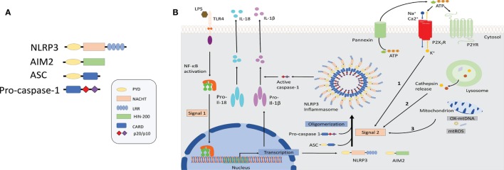
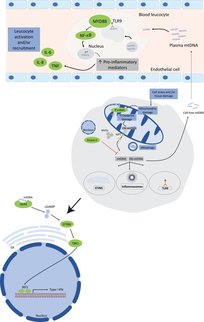
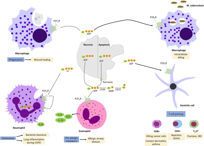
Over the recent years, much has been unraveled about the pro-inflammatory properties of various mitochondrial molecules once they are leaving the mitochondrial compartment. On entering the cytoplasm or the extracellular space, mitochondrial DAMPs (also known as mitochondrial alarmins) can become pro-inflammatory and initiate innate and adaptive immune responses by activating cell surface and intracellular receptors. Current evidence indicates that uncontrolled and excessive release of mitochondrial DAMPs is associated with severity, has prognosis value in human diseases, and contributes to the dysregulated process observed in numerous inflammatory and autoimmune conditions, as well as in ischemic heart disease and cancer. Herein, we review that the expanding research field of mitochondrial DAMPs in innate immune responses and the current knowledge on the association between mitochondrial DAMPs and human diseases.

Keywords: alarmins; damage-associated molecular pattern; inflammation; mitochondria; pro-inflammatory cytokines; sterile inflammation.


Figures

Figure 1
Figure 1
Schematic illustration of selected inflammasome components and mechanism of NLRP3 activation. (A) Shown here is the composition of NLRP3 and absent in melanoma-2 inflammasome sensors as well as the components ASC and caspase-1. (B) NLRP3 inflammasomes activation involves a two-step process: priming and assembly. Signal 1 (priming) is provided by nuclear factor-κB-dependent transcription of pro-interleuking-1β and NLRP3, either through the activation of TLRs or nucleotide-binding oligomerization domain 2 (NOD2) by microbial molecules or endogenous cytokines. The second signal is provided by stimuli that specifically activate NLRP3 and lead to NLRP3 oligomerization, caspase-1 activation followed by the maturation and release of IL-1β and IL-18. Three separate phenomena had been associated with NLRP3 activation. Event 1: ATP mediated ionic flux and intracellular potassium depletion mediated by ATP binding to ligand-gated ion channels P2X7R. Event 2: cathepsin release following destabilization of lysosomal membrane by sterile particulates, such as silica, asbestos, and cholesterol crystals. Event 3: generation of ROS and cytoplasmic release of mitochondrial DNA following major cellular stress and mitochondrial damage. Abbreviations: ATP, adenosine triphosphate; AIM2, absent in melanoma-2; ASC, apoptosis-associated speck-lick protein containing a CARD; CARD, caspase-activation and recruitment domain; HIN-200, hematopoietic interferon-inducible nuclear proteins with a 200-amino-acid repeat; IL-1β/IL-18, interleukin 1β/18; LPS, lipopolysaccharide; LRR, leucine-rich repeats; NBD/NACHT, nuclear binding domain; NF-κB, nuclear factor-κB; NLRP3, NLR family pyrin domain-containing; NLR, nucleotide-binding domain and leucine-rich repeat receptors; NOD2, nucleotide-binding oligomerization domain 2; PYD, pyrin domain; ROS, reactive oxygen species; TLRs, toll-like receptors.
Figure 2
Figure 2
Mitochondrial DNA (mtDNA) mediates a pro-inflammatory response by interacting with TLR9, cytosolic inflammasomes, and type I interferon response. Tissue injury or cellular damage can cause mitochondrial dysfunction resulting in oxidative damage with increased mitochondrial reactive oxygen species (ROS) production and mtDNA oxidation. Oxidized mtDNA will be released into the cytosol and then to the extracellular milieu by various mechanisms including the transport in mitochondrial derived vesicles (MVDs) or through mitochondrial permeability transition pores (MPT). The whole damaged mitochondria can also be recycled through the mitophagy process. In the plasma, mtDNA activates TLR9-mediated signaling pathway in circulating neutrophils resulting in increased production of pro-inflammatory mediators, such as tumor necrosis factor (TNF), interleukin-6 (IL-6), and adhesion molecules. Cytosolic mtDNA can also engage and activate the NOD, leucine-rich repeats, and pyrin domain-containing protein 3 (NLRP3) inflammasome or absent in melanoma 2 inflammasome and trigger a pro-inflammatory response in cells primed with first signal in inflammasome activation through the nuclear factor (NF)-κB pathway. Finally, cytosolic mtDNA can also be recognized by and engage the cyclic GMP-AMP synthase (cGAS). Once activated cGAS triggers conformational changes of the endoplasmic reticulum-resident protein stimulator of interferon genes (STING), which engages TANK-binding kinase 1 to activate interferon regulatory factor 3 (IRF3) and/or IRF7 to stimulate transcription of type I interferons (IFNs) genes.
Figure 3
Figure 3
P2Y2R and P2X7R signaling during injury and inflammation. Adenosine triphosphate (ATP) can be released by necrotic cells or apoptotic cells though cytoplasmic channel, pannexin 1. When engaging P2Y2R, released ATP functions as “find me signal” for macrophages phagocytosis of injured cells and promotes so wound healing. P2Y2R signaling contributes also to bacterial clearance by stimulating neutrophils chemotaxis. However, excess P2Y2R signaling in neutrophils or eosinophils contributes, respectively, to chronic lung inflammation and allergic airway disease by promoting the release of pro-allergic mediators. P2X7R signaling promotes the killing of intracellular bacteria such as Mycobacter Tuberculosis. In dendritic cells, P2X7R signaling promotes T-cell priming which has been implicated in allergic diseases, graft-versus-host disease (GVH) as well as in psoriasis and inflammatory bowel disease.

References

    1. Matzinger P. The danger model: a renewed sense of self. Science (2002) 296:301–5.10.1126/science.1071059 - DOI - PubMed
    1. Chen GY, Nunez G. Sterile inflammation: sensing and reacting to damage. Nat Rev Immunol (2010) 10:826–37.10.1038/nri2873 - DOI - PMC - PubMed
    1. Scaffidi P, Misteli T, Bianchi ME. Release of chromatin protein HMGB1 by necrotic cells triggers inflammation. Nature (2002) 418:191–5.10.1038/nature00858 - DOI - PubMed
    1. Quintana FJ, Cohen IR. Heat shock proteins as endogenous adjuvants in sterile and septic inflammation. J Immunol (2005) 175:2777–82.10.4049/jimmunol.175.5.2777 - DOI - PubMed
    1. Schroder K, Tschopp J. The inflammasomes. Cell (2010) 140:821–32.10.1016/j.cell.2010.01.040 - DOI - PubMed

Publication types