Review
. 2018 May 4:9:158.
doi: 10.3389/fgene.2018.00158. eCollection 2018.

Intrinsic Disorder and Posttranslational Modifications: The Darker Side of the Biological Dark Matter

Affiliations
Review

Intrinsic Disorder and Posttranslational Modifications: The Darker Side of the Biological Dark Matter

April L Darling et al. Front Genet. .

Abstract

Intrinsically disordered proteins (IDPs) and intrinsically disordered protein regions (IDPRs) are functional proteins and domains that devoid stable secondary and/or tertiary structure. IDPs/IDPRs are abundantly present in various proteomes, where they are involved in regulation, signaling, and control, thereby serving as crucial regulators of various cellular processes. Various mechanisms are utilized to tightly regulate and modulate biological functions, structural properties, cellular levels, and localization of these important controllers. Among these regulatory mechanisms are precisely controlled degradation and different posttranslational modifications (PTMs). Many normal cellular processes are associated with the presence of the right amounts of precisely activated IDPs at right places and in right time. However, wrecked regulation of IDPs/IDPRs might be associated with various human maladies, ranging from cancer and neurodegeneration to cardiovascular disease and diabetes. Pathogenic transformations of IDPs/IDPRs are often triggered by altered PTMs. This review considers some of the aspects of IDPs/IDPRs and their normal and aberrant regulation by PTMs.

Keywords: intrinsically disordered protein regions; intrinsically disordered proteins; multifunctional proteins; posstranslational modifications; protein–protein interaction (PPI).


Figures

FIGURE 1
FIGURE 1
Abundance of intrinsic disorder in various proteomes.
FIGURE 2
FIGURE 2
Involvement of intrinsic disorder in protein function. Note that the classical structure-function paradigm cannot describe many of the function proteins perform.
FIGURE 3
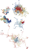
FIGURE 3
Illustrative examples of proteins with different levels of global disorder. (A) Ordered protein: NMR solution structure of bovine ubiquitin (PDB ID: 1V81) (Kitahara et al., 2005). (B) Protein with the collapsed (molten globule-like) disorder: NMR solution structure of Ubiquitin-like domain of NFATC2IP (PDB ID: 2JXX). (C) Protein with semi-extended (pre-molten globule-like) disorder: NMR solution structure of the domain II from the blood-stage malarial protein, apical membrane antigen 1 (PDB ID: 1YXE) (Feng et al., 2005). (D) Small protein with extended (coil-like) disorder: sunflower trypsin inhibitor 1 (SFTI-1) (PDB ID: 2AB9) (Mulvenna et al., 2005). (E) Large protein with extended (coil-like) disorder: NMR solution structure of Air2p protein, which is the key component of the Trf4/5p-Air1/2p-Mtr4p polyadenylation complex (TRAMP) (PDB ID: 2LLI) (Holub et al., 2012).
FIGURE 4
FIGURE 4
Disordered nature of the PTM sites in target proteins. (A) Crystal structure of a complex between a 20-amino acid peptide derived from the heat stable protein kinase inhibitor (PKIα) and the catalytic subunit of cyclic AMP-dependent protein kinase (cAPK) (PDB ID: 2CPK) (Knighton et al., 1991). (B) Crystal structure of a complex between a 23 amino acid cell-permeable peptide and a protein Ser/Thr phosphatase-1 (PDB ID: 4G9J) (Chatterjee J. et al., 2012). (C) Crystal structure of a complex between polypeptide N-acetylgalactosaminyltransferase 2 and a 13 amino acid peptide EA2 (PDB ID: 2FFU) (Abraham and Podell, 1981). (D) Crystal structure of a complex between human mitochondrial NAD-dependent deacetylase sirtuin-3 and a 12 amino acid peptide derived from mitochondrial acetyl-coenzyme A synthetase 2-like (PDB ID: 3GLT) (Jin et al., 2009). (E) Crystal structure of a complex between a protein arginine N-methyltransferase 1 and a 19 amino acid substrate peptide (PDB ID: 1OR8) (Zhang and Cheng, 2003).

References

    1. Abraham G. N., Podell D. N. (1981). Pyroglutamic acid. Non-metabolic formation, function in proteins and peptides, and characteristics of the enzymes effecting its removal. Mol. Cell. Biochem. 38 181–190. 10.1007/BF00235695 - DOI - PubMed
    1. Antz C., Geyer M., Fakler B., Schott M. K., Guy H. R., Frank R., et al. (1997). NMR structure of inactivation gates from mammalian voltage-dependent potassium channels. Nature 385 272–275. 10.1038/385272a0 - DOI - PubMed
    1. Apweiler R., Hermjakob H., Sharon N. (1999). On the frequency of protein glycosylation, as deduced from analysis of the SWISS-PROT database. Biochim. Biophys. Acta 1473 4–8. 10.1016/S0304-4165(99)00165-8 - DOI - PubMed
    1. Ardito F., Giuliani M., Perrone D., Troiano G., Lo Muzio L. (2017). The crucial role of protein phosphorylation in cell signaling and its use as targeted therapy (Review). Int. J. Mol. Med. 40 271–280. 10.3892/ijmm.2017.3036 - DOI - PMC - PubMed
    1. Armstrong C. M., Bezanilla F. (1977). Inactivation of the sodium channel. II. Gating current experiments. J. Gen. Physiol. 70 567–590. 10.1085/jgp.70.5.567 - DOI - PMC - PubMed

LinkOut - more resources