. 2018 Apr 3;9(18):4285-4298.
doi: 10.1039/c8sc00804c. eCollection 2018 May 14.

Counting charges on membrane-bound peptides

Affiliations

Counting charges on membrane-bound peptides

Alicia C McGeachy et al. Chem Sci. .

Abstract

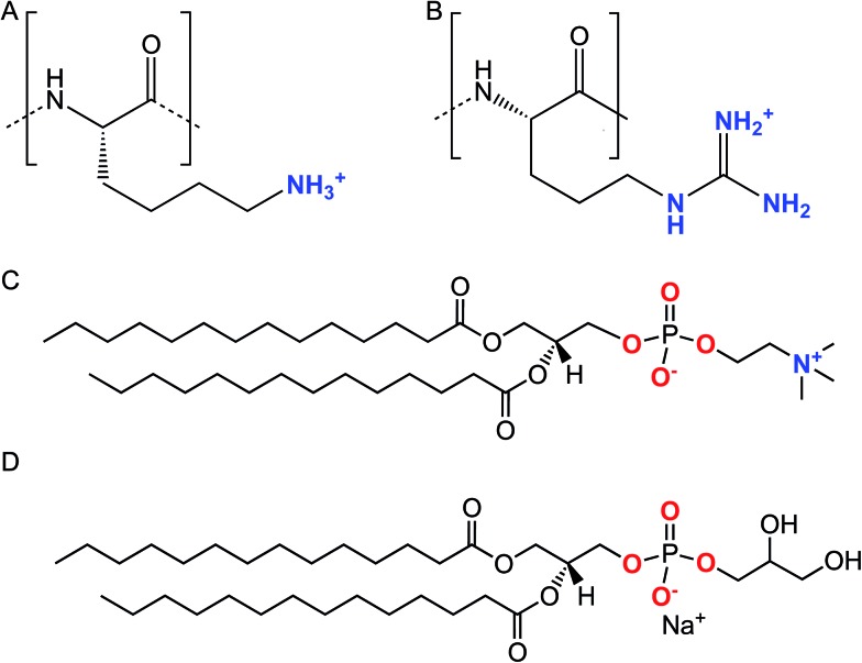
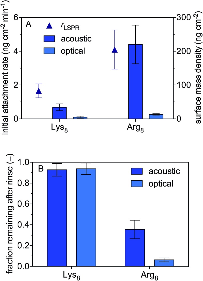
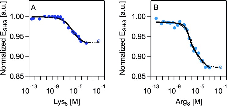
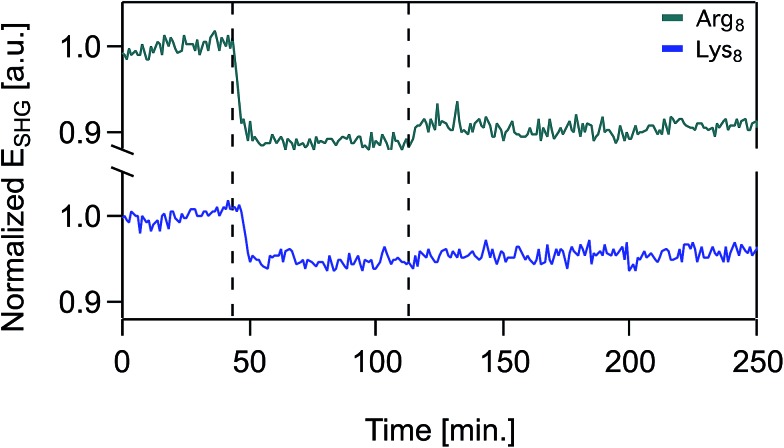
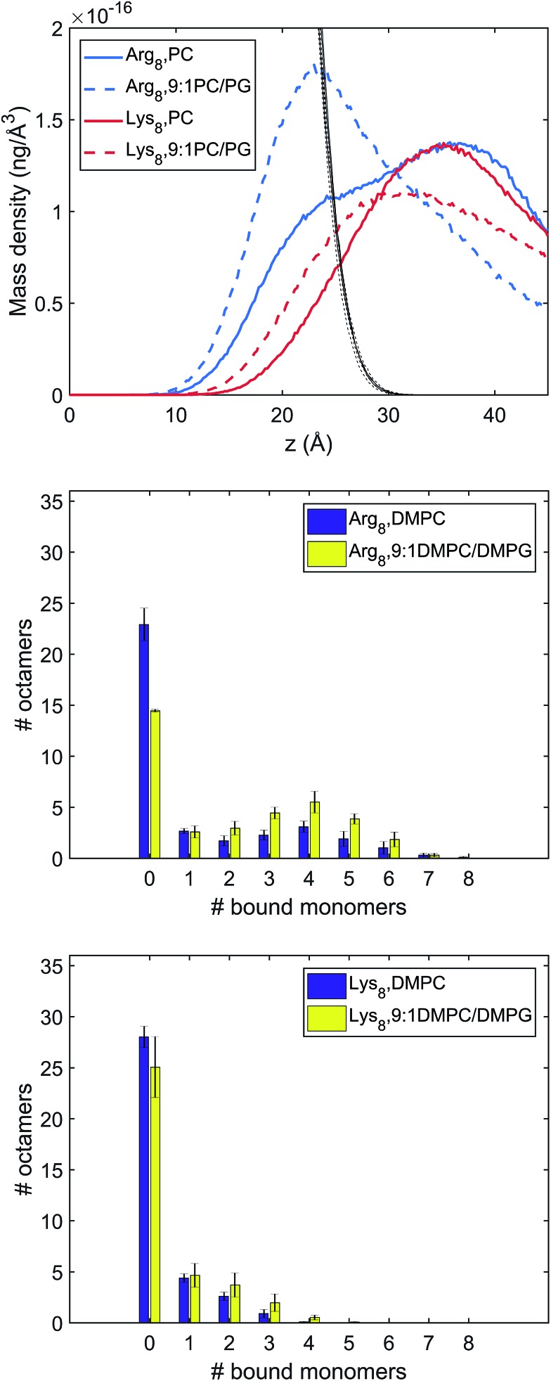
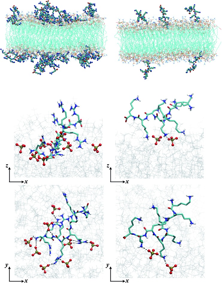
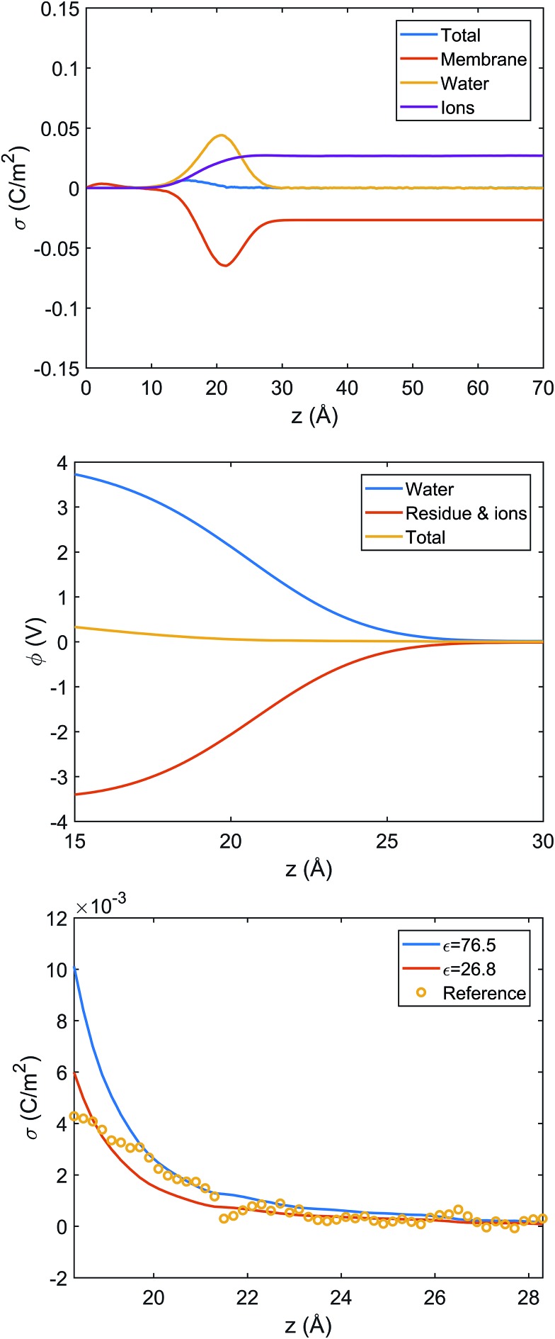
Quantifying the number of charges on peptides bound to interfaces requires reliable estimates of (i) surface coverage and (ii) surface charge, both of which are notoriously difficult parameters to obtain, especially at solid/water interfaces. Here, we report the thermodynamics and electrostatics governing the interactions of l-lysine and l-arginine octamers (Lys8 and Arg8) with supported lipid bilayers prepared from a 9 : 1 mixture of 1,2-dimyristoyl-sn-glycero-3-phosphocholine (DMPC) and 1,2-dimyristoyl-sn-glycero-3-phospho-(1'-rac-glycerol) (sodium salt) (DMPG) from second harmonic generation (SHG) spectroscopy, quartz crystal microbalance with dissipation monitoring (QCM-D) and nanoplasmonic sensing (NPS) mass measurements, and atomistic simulations. The combined SHG/QCM-D/NPS approach provides interfacial charge density estimates from mean field theory for the attached peptides that are smaller by a factor of approximately two (0.12 ± 0.03 C m-2 for Lys8 and 0.10 ± 0.02 C m-2 for Arg8) relative to poly-l-lysine and poly-l-arginine. These results, along with atomistic simulations, indicate that the surface charge density of the supported lipid bilayer is neutralized by the attached cationic peptides. Moreover, the number of charges associated with each attached peptide is commensurate with those found in solution; that is, Lys8 and Arg8 are fully ionized when attached to the bilayer. Computer simulations indicate Lys8 is more likely than Arg8 to "stand-up" on the surface, interacting with lipid headgroups through one or two sidechains while Arg8 is more likely to assume a "buried" conformation, interacting with the bilayer through up to six sidechains. Analysis of electrostatic potential and charge distribution from atomistic simulations suggests that the Gouy-Chapman model, which is widely used for mapping surface potential to surface charge, is semi-quantitatively valid; despite considerable orientational preference of interfacial water, the apparent dielectric constant for the interfacial solvent is about 30, due to the thermal fluctuation of the lipid-water interface.


Figures

Scheme 1
Scheme 1. Chemical structures of the lysine (A) and arginine (B) repeat units in Lys8 and Arg8. Chemical structures of DMPC (C) and DMPG (D).
Fig. 1
Fig. 1. Attachment of octamers of lysine (Lys8) and arginine (Arg8) to supported lipid bilayers formed from 9 : 1 DMPC/DMPG. (A) Initial attachment rates and maximum acoustic and optical surface mass densities. The initial attachment rates were based on optical masses calculated from localized surface plasmon resonance data. (B) Acoustic and optical surface mass densities after 10 min rinse with oligomer-free solution. Solutions were 0.10 M NaCl buffered to pH 7.4 with 0.01 M Tris. Error bars represent one standard deviation of triplicate measurements.
Fig. 2
Fig. 2. Normalized SHG E-field as a function of polymer concentration, in molarity, at 0.1 M NaCl, 0.01 M Tris, pH 7.4 for (A) Lys8 and (B) Arg8. Data sets include an extrapolated data point (shown as an open circle) that is the average of the last three measured data points. SHG E-field is normalized to the signal intensity associated with the supported lipid bilayer formed from 9 : 1 DMPC/DMPG prior to exposure to oligomers. Each individual adsorption isotherm is shown with the corresponding fit with the combined Hill/Gouy–Chapman equation applied to the acquired data (black solid line) and complete data set with extrapolated data point (dashed black line). See main text for further discussion.
Fig. 3
Fig. 3. Normalized SHG E-field as a function of time in the presence of supported lipid bilayers formed from 9 : 1 DMPC/DMPG for 50 μM Arg8 (top trace, green), 50 μM Lys8 (bottom trace, blue) at 0.1 M NaCl, 0.01 M Tris, pH 7.4. At t = 0, the supported lipid bilayer is unperturbed and the SHG signal is monitored at 0.1 M NaCl. At t = 43 min, oligomer solution is introduced into the flow cell and at t = 112 min the flow cell is rinsed with oligomer-free solution composed of otherwise identical composition.
Fig. 4
Fig. 4. Characterization of different binding modes of Arg8 and Lys8 to the lipid membrane. Top: Mass density of peptide atoms along z (the membrane normal); the black lines indicate the mass density of lipid atoms. Middle and bottom panels: Distribution of the number of amino acid side chains bound to the lipid membrane (# of bound monomers) for the 36 copies of peptides in the simulation cell. The middle panel is for Arg8 and the bottom panel for Lys8. Evidently, Arg8 is observed to interact with the membrane through multiple sidechains, especially in the presence of anionic lipids; by contrast, Lys8 interact with the membrane with a small (1–3) number of sidechains.
Fig. 5
Fig. 5. Snapshots from MD simulations (Arg8/Lys8 interacting with a 9 : 1 DMPC/DMPG bilayer) illustrate the different binding modes of the peptides. The left column is for Arg8, and right column for Lys8. The top two rows are sideviews (showing multiple peptides and a single peptide, respectively), which illustrate that due to the different numbers of sidechains interacting with the lipids, Lys8 peptides tend to point into the solution, while Arg8 peptides lay closer to the membrane; these trends are also illustrated by the mass density distributions (Fig. 4, top panel). The bottom row contains the top view of close-ups of the binding interactions; while Arg8 are engaged with multiple phosphate groups (those within 3 Å from Arg8 are shown in CPK), only a few lipid phosphate groups interact with the Lys sidechains.
Fig. 6
Fig. 6. Analysis of charge distribution and electrostatic potential near the lipid/water interface for a 9 : 1 DMPC/DMPG bilayer; corresponding analyses for the bilayer with bound Arg8 and Lys8 peptides are discussed in the ESI. Top: Integrated charge density from the center of the bilayer (z = 0), , where ρ(z′) is the charge density binned along z (the direction of the membrane normal) averaged over snapshots from MD simulations; note the significant contribution from interfacial water due to orientational preference. Middle: Electrostatic potential computed based on the charge density from MD simulations (eqn (13) in ESI†) illustrates a strong compensation between interfacial water and other components (lipids and salt ions). Bottom: Surface charge density computed with Grahame's equation and the electrostatic potential from MD simulations using different values of dielectric constant for the interfacial solvent. The open circles indicate integrated charge density from MD simulations (i.e., the top panel). Since the precise location of the interface is not straightforward to determine, calculations based on the Grahame's equation are done for a series of z values near the location of the phosphate groups (z ∼ 20 Å). See ESI for additional discussions.

References

    1. Salay L. C., Petri D. F. S., Nakaie C. R., Schreier S. Biophys. Chem. 2015;207:128–134. - PubMed
    1. Wilhelm M. J., Gh M. S., Dai H. L. Biochemistry. 2015;54:4427–4430. - PubMed
    1. Gong Y., Andina D., Nahar S., Leroux J. C., Gauthier M. A. Chem. Sci. 2017;8:4082–4086. - PMC - PubMed
    1. Nystrom L., Nordstrom R., Bramhill J., Saunders B. R., Alvarez-Asencio R., Rutland M. W., Malmsten M. Biomacromolecules. 2016;17:669–678. - PubMed
    1. Epand R. M., Epand R. F. Biochim. Biophys. Acta, Biomembr. 2009;1788:289–294. - PubMed

LinkOut - more resources