Case Reports
. 2018 Mar 26:2018:1370701.
doi: 10.1155/2018/1370701. eCollection 2018.

Spindle Cell Hemangioma in the Mucosa of the Upper Lip: A Case Report and Review of the Literature

Affiliations
Case Reports

Spindle Cell Hemangioma in the Mucosa of the Upper Lip: A Case Report and Review of the Literature

Kazuhiro Murakami et al. Case Rep Dent. .

Abstract

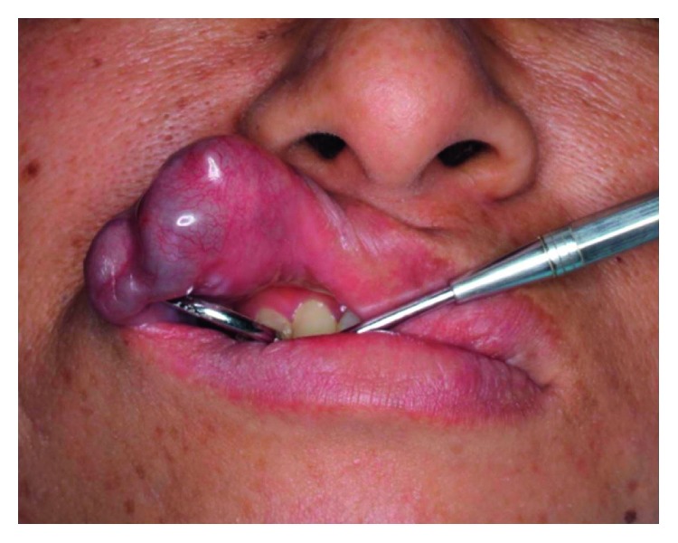
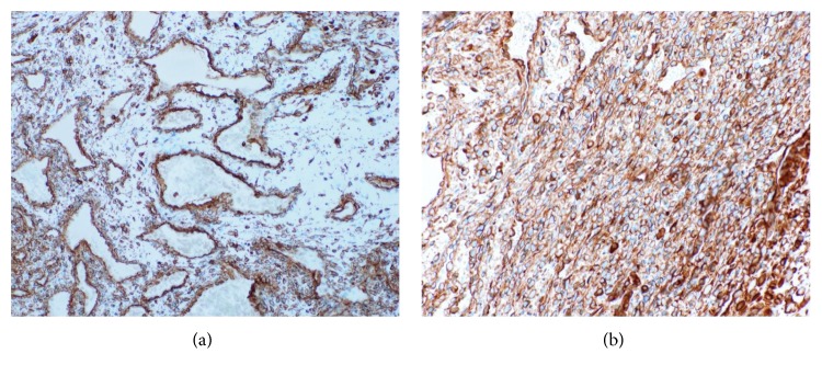
Spindle cell hemangioma (SCH) is a unique benign vascular lesion. We present a case of SCH in the upper lip of a 41-year-old woman. A submucosal nodular mass 30 × 20 mm in size was observed in the left upper lip. The mass developed 5 years earlier and enlarged after repeated ethanol injections. The mass was elastic firm, mobile, bluish in color, and well demarcated in magnetic resonance imaging. Under the clinical diagnosis of hemangioma, surgical excision was performed under local anesthesia. Microscopically, the lesion was composed of irregular cavernous spaces and multiple solid cellular areas. Cavernous spaces were filled with a mix of erythrocytes and organizing thrombi. The solid areas showed proliferation of spindle-shaped cells arranged haphazardly or in short interlacing fascicles. Immunohistochemically, most cells strongly reacted with vimentin. CD31, CD34, factor VIII, smooth muscle actin, and Wilms tumor-1 reacted with endothelial cells lining the cavernous spaces. The cells within solid areas consisted of mixed cell population with variable reaction for the markers except for factor VIII. From these findings, the diagnosis of SCH was made. Two years after surgery, no recurrence was noted. A review of SCH in the head and neck region is made.


Figures

Figure 1
Figure 1
Clinical finding before surgery.
Figure 2
Figure 2
MRI findings. (a) T1-weighted image. (b) T2-weighted image.
Figure 3
Figure 3
Excised tumor: lateral view.
Figure 4
Figure 4
Clinical finding 6 months after surgery.
Figure 5
Figure 5
(a) Well-defined submucosal mass with cavernous spaces, solid areas, and number of thrombi. (hematoxylin and eosin stain, original magnification ×5). (b) Irregular cavernous spaces lined by flat endothelial cells. (hematoxylin and eosin stain, original magnification ×100). (c) The spindle-shaped cells in solid areas. (hematoxylin and eosin stain, original magnification ×100).
Figure 6
Figure 6
(a) Cavernous area (vimentin, original magnification ×100). Most endothelial cells around blood vessels in cavernous area strongly reacted. (b) Solid area (vimentin, original magnification ×200). Spindle cells and epithelioid in solid area are strongly positive.
Figure 7
Figure 7
(a) Cavernous area (CD34, original magnification ×100). Most endothelial cells around vessels in cavernous space are positive. (b) Solid area (CD34, original magnification ×200). Spindle cells in solid area are focally positive.
Figure 8
Figure 8
(a) Cavernous area (SMA, original magnification ×100). Most endothelial cells around vessels are positive. (b) Solid area (SMA, original magnification ×200). Spindle cells in solid area are focally positive.
Figure 9
Figure 9
(a) Cavernous area (WT-1, original magnification ×100). Endothelial cells are strongly positive in cavernous area. (b) Solid area (WT-1, original magnification ×200). Spindle cells in solid area are focally positive.

References

    1. Tosios K., Gouveris I., Sklavounou A., Koutlas I. G. Spindle cell hemangioma (hemangioendothelioma) of the head and neck: case report of an unusual (or underdiagnosed) tumor. Oral Surgery, Oral Medicine, Oral Pathology, Oral Radiology, and Endodontology. 2008;105:216–221. doi: 10.1016/j.tripleo.2007.03.005. - DOI - PubMed
    1. Scott G. A., Rosai J. Spindle cell hemangioendothelioma: report of seven additional cases of a recently described vascular neoplasm. American Journal of Dermatopathology. 1988;10:281–288. doi: 10.1097/00000372-198808000-00001. - DOI - PubMed
    1. Tosios K., Koutlas I. G., Kapranos N., Papanicolaou S. I. Spindel-cell hemangioendothelioma of the oral cavity. A case report. Journal of Oral Pathology and Medicine. 1995;24:379–382. doi: 10.1111/j.1600-0714.1995.tb01203.x. - DOI - PubMed
    1. Baron J. A., Raines J., Bangert J., Hansen R. C. Persistent nodule on the nose. Archives of Dermatology. 2002;138:259–264. doi: 10.1001/archderm.138.2.259-b. - DOI - PubMed
    1. Ide F., Obara K., Enatsu K., Mishima K., Saito I. Rare vascular proliferations of the oral mucosa. Oral Surgery, Oral Medicine, Oral Pathology, Oral Radiology, and Endodontology. 2004;97:75–78. doi: 10.1016/j.tripleo.2003.08.030. - DOI - PubMed

Publication types

LinkOut - more resources