Case Reports
. 2018 May 2;8(2):e2018022.
doi: 10.4322/acr.2018.022. eCollection 2018 Apr-Jun.

Congenital pulmonary airway malformation

Affiliations
Case Reports

Congenital pulmonary airway malformation

Wlamir Pestana Ursini et al. Autops Case Rep. .

Abstract

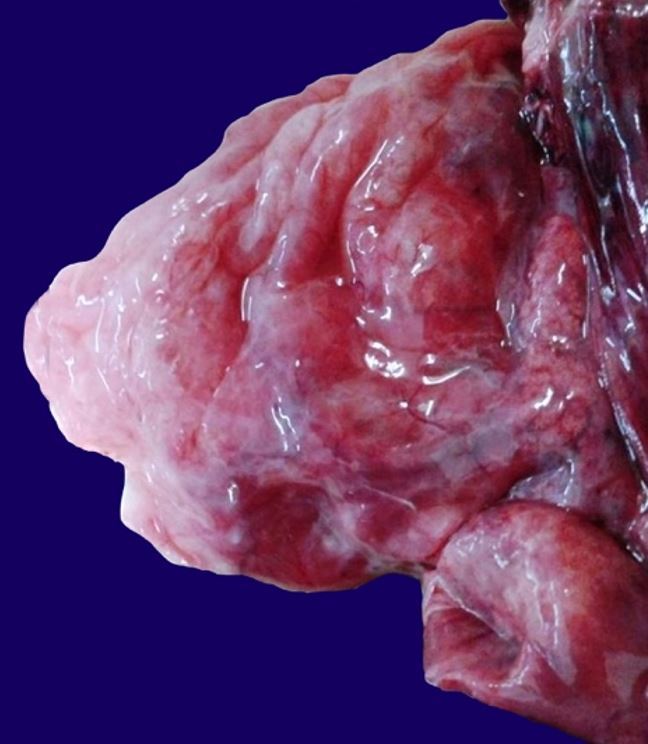
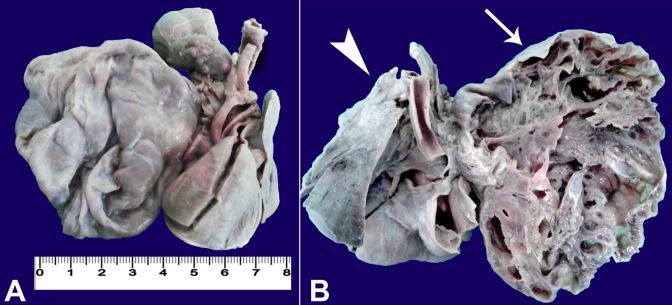
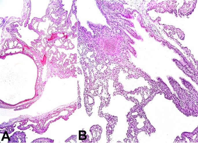
Congenital pulmonary airway malformation (CPAM) is a developmental malformation of the lower respiratory tract. We report the case of a male newborn diagnosed with cystic lung disease during prenatal ultrasound. A cesarean section was performed at the 32nd gestational week because of premature rupture of the membranes, and soon after the delivery the newborn developed respiratory failure and died. The aim of this study is to report an autopsy case because of its rarity, and to briefly discuss the CPAM subtypes and differential diagnosis of cystic lung diseases of childhood.

Keywords: Autopsy; Cystic Adenomatoid Malformation of Lung, Congenital; Newborn; Respiratory Distress Syndrome.


Conflict of interest statement

Conflict of interest: None

Figures

Figure 1
Figure 1. Gross view of the hyperinflated right lung with multiple cysts.
Figure 2
Figure 2. A – Front view of the thoracic organs after formalin fixation; B – Posterior view of the thoracic organs. Note the right lung with numerous contiguous cystic formations outlined by thin walls with honeycomb appearance (arrow) and the immature left lung with the two lobes (arrowhead).
Figure 3
Figure 3. Photomicrograph of the right lung revealing in A – Cystic dilated bronchiolar structures (H&E, 200X); and B – Polypoid projections of the mucosa and rare cartilage foci in the bronchial wall (H&E, 200X).
Figure 4
Figure 4. A – Photomicrograph of the right lung revealing clusters of mucinous glands (PAS, 200X); and B – Presence of the goblet cells (PAS, 400X).

References

    1. Andrade CF, Ferreira HPC, Fischer GB. Congenital lung malformations. J Bras Pneumol. 2011;37(2):259-71. 10.1590/S1806-37132011000200017. - DOI - PubMed
    1. Reis AR, Ribeiro FB, Schultz R. Congenital cystic adenomatoid malformation type I. Autops Case Rep. 2015;5(3):21-6. 10.4322/acr.2015.019. - DOI - PMC - PubMed
    1. Chong Y, Rhee YJ, Han SJ, Cho HJ, Kang SK, Kang MW. Life-threatening congenital cystic adenomatoid malformation in the premature neonate. Korean J Thorac Cardiovasc Surg. 2016;49(3):210-3. 10.5090/kjtcs.2016.49.3.210. - DOI - PMC - PubMed
    1. Sfakianaki AK, Copel JA. Congenital cystic lesions of the lung: congenital cystic adenomatoid malformation and bronchopulmonary sequestration. Rev Obstet Gynecol. 2012;5(2):85-93. - PMC - PubMed
    1. David M, Lamas-Pinheiro R, Henriques-Coelho T. Prenatal and postnatal management of congenital pulmonary airway malformation. Neonatology. 2016;110(2):101-15. 10.1159/000440894. - DOI - PubMed

Publication types