. 2018 Aug;75(8):339-351.
doi: 10.1002/cm.21454.

Nonmuscle myosin II in cardiac and skeletal muscle cells

Affiliations

Nonmuscle myosin II in cardiac and skeletal muscle cells

Jushuo Wang et al. Cytoskeleton (Hoboken). 2018 Aug.

Abstract

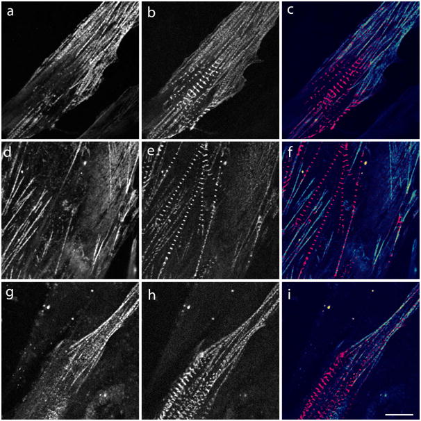
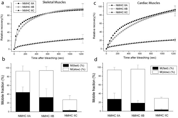
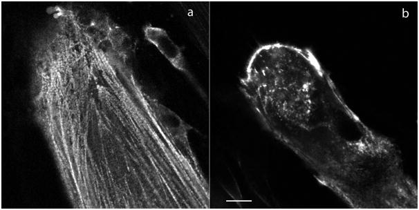
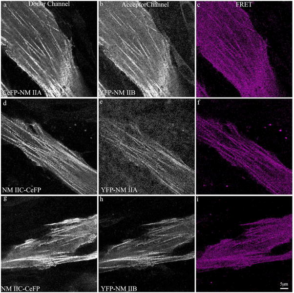
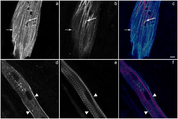
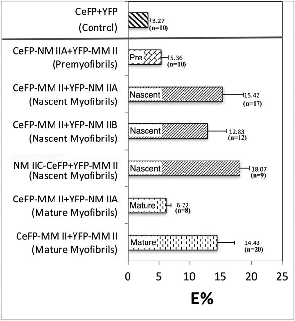
De novo assembly of contractile myofibrils begins with the formation of premyofibrils where filaments of non-muscle myosin (NM II), and actin organize in sarcomeric patterns with Z-Bodies containing muscle-specific alpha-actinin. Interactions of muscle specific myosin (MM II) with NM II occur in a nascent myofibril stage that precedes the assembly of mature myofibrils. By the final stage of myofibrillogenesis, the only myosin II present in the mature myofibrils is MM II. In this current study of myofibril assembly, the three vertebrate isoforms of NM II (A, B, and C) and sarcomeric alpha-actinin, ligated to GFP family proteins, were coexpressed in avian embryonic skeletal and cardiac muscle cells. Each isoform of NM II localized only in the mini-A-Bands of premyofibrils and nascent myofibrils. There was no evidence of localization of NM II in Z-Bodies of premyofibrils and nascent myofibrils or in Z-Bands of mature myofibrils. Fluorescence Recovery After Photobleaching (FRAP) experiments indicated similar exchange rates in premyofibrils for NM II isoforms A and B, whereas the IIC isoform was significantly less dynamic. Fluorescence Resonance Energy Transfer (FRET) measurements of colocalized fluorescent pairs of different NM II isoforms yielded signals similar to identical pairs, indicating copolymerization of the different NM II pairs. The role of NM II may reside in establishing the future sarcomere pattern in mature myofibrils by binding to the oppositely polarized actin filaments that extend between pairs of Z-Bodies along premyofibrils prior to their transformation into mature myofibrils.

Keywords: FRAP; FRET; Myofibrillogenesis; mature myofibril; nascent myofibril; non-muscle myosin II isoforms; premyofibril; sarcomere.


Figures

Figure 1
Figure 1. Expression of each of the three isoforms of nonmuscle myosin II (NM II) in live myotubes leads to similar patterns of incorporation in premyofibrils
Premyofibrils in the growing ends of live quail myotubes were recorded two days after cotransfections with plasmids encoding GFP and non-muscle myosin II isoforms (a, GFP NM IIA; d, GFP-NM IIB; g, NM IIC-GFP; cyan in c, f, i), together with sarcomeric alpha-actinin-CeFP (b, e, h, magenta in c, f, i). Each isoform of non-muscle myosin: IIA (a), IIB (d), and IIC (g) localized in premyofibrils in small bands that alternated with bands of alpha-actinin: merged images (c, f, i). Boxed areas are shown enlarged in the merged column. Bar = 10 microns (a-i). Bar = 2 microns (inset in c, f, i).
Figure 2
Figure 2. Expression of each isoform of NM II in older myotubes leads to their localization in thin premyofibrils along the myotube membrane adjacent to mature myofibrils
Each isoform of non-muscle myosin II coupled to GFP incorporated in a few thin premyofibrils adjacent to the membrane surrounding mature myofibrils in the middle of myotubes: (a) IIA; (d) IIB; (g) IIC (cyan in c, f, i). alpha-actinin-CeFP localization in Z-bands marks mature myofibrils in the myotube center (b, e, h; magenta in c, f, i). Note that none of the three fluorescently labeled isoforms of NM II is localized in Z-Bands (c, f, i). Bar = 10 microns.
Figure 3
Figure 3. Spatially distinct patterns of localization of NM IIA and sarcomeric alpha-actinin are illustrated in axial views, XY, XZ, and YZ, of a myotube cotransfected with GFP-NM IIA and sarcomeric alpha-actinin-CeFP
Three views of a confocal image of the same live myotube show NM IIA (a, cyan in c) localized around the periphery of the myotube, and absent from the center of the myotube where alpha-actinin is concentrated in Z-Bands (b, magenta in c). Merged view (c) indicates the lack of any overlap between Z-Bands (magenta) and NM II (cyan) in mature myofibrils. Bar = 10 microns.
Figure 4
Figure 4. Overexpression of non-muscle myosin IIA in a myotube results in its localization in A-Bands and in aggregates
Four days after myotubes were cotransfected with (a) alpha-actinin-CeFP and (b) GFP-NM IIA, they were fixed and stained with muscle-specific myosin II (MM II) antibody, F-59 (c). Arrowheads (a, b, c; images enlarged in d to i) mark the positions of three Z-Bands (blue in g and i). (b, e, green in g) Single bands of GFP-NM IIA localize between the Z-bands (arrowheads). (c, f, i) F-59 antibody staining shows muscle myosin heavy chain (MM II) localized in a doublet pattern in A-Bands (red in h and i) between the Z-Bands (arrowheads). Note that even when overexpressed, NM IIA did not colocalize with alpha-actinin in Z-Bands in mature myofibrils (b, e, g). The magnified views of NM IIA (e, green in g and h) and MM II (f, red in h and i) indicate that the bands of NM IIA formed in the center of the MM II doublet in the A-Bands. Arrows in (b) point out nonsarcomeric aggregates of nonmuscle myosin IIA. Bar = 10 microns (a-c); 2 microns (d-i).
Figure 5
Figure 5. Expression of each of the three isoforms of nonmuscle myosin II (NM II) in living embryonic chick cardiomyocytes leads to similar patterns of incorporation in premyofibrils
Premyofibrils are detected two days after cotransfections by their fluorescent non-muscle myosin II isoforms. (a) GFP-non-muscle myosin IIA; cyan in (c). (d) GFP-non-muscle myosin IIB; cyan in (f). (g) non-muscle myosin IIC-GFP; cyan in (i). (b, e, h) Mature myofibrils are detected in these same cells by the localization of sarcomeric alpha-actinin-CeFP in their Z-Bands (b, e, h; magenta in c, f, i). Note that none of the three different isoforms of non-muscle myosin II are localized in the Z-Bands (c, f. i). Bar = 10 microns.
Figure 6
Figure 6. Fluorescence Recovery after Photobleaching (FRAP) compares the dynamic exchange and mobile fractions of NM II isoforms in premyofibrils in skeletal and cardiac myocytes
In each muscle type, recovery curves and mobile fractions calculated after photobleaching show similar values for NM IIA and IIB in skeletal muscle (a, b) and in cardiomyocytes (c, d). Whereas in both types of muscle, IIC recovery occurred much more slowly (a, b and c, d).
Figure 7
Figure 7. Expression of NM IIC-GFP in myotubes leads initially to its incorporation in a banded pattern in premyofibrils. Several days after transfection, however, premyofibrils disassemble
(a) NM IIC-GFP assembled in premyofibrils in the flat, spreading end of a myotube two days after transfection. (b) Four days after transfection of NM IIC-GFP, premyofibrils had disassembled. Bar = 10 microns.
Figure 8
Figure 8. SE-FRET analysis suggests that isoforms of NM II copolymerize in premyofibrils in live quail myotubes
Pairs of plasmids encoding CeFP or YFP and different isoforms of NM II were cotransfected in quail myotubes: (a, b) CeFP-NM IIA and YFP-IIB pair; (d, e) NM IIC-CeFP and YFP-IIA pair; and (g, h) NM IIC-CeFP and YFP-IIB pair. FRET signals were acquired in three channels: donor channel (a, d, g for CeFP), acceptor channel (b, e, h for YFP) and the resultant FRET channel (c, f, i). FRET efficiencies were calculated from these three-channel images and included in Table 2. Bar = 5 μm.
Figure 9
Figure 9. NMII and MM II colocalize in nascent myofibrils early in myofibril assembly in living myotubes
Cultures of quail myotubes were cotransfected with CeFP-NM IIA (a, cyan in c, f), and YFP-MM II (b, magenta in c, f). NM IIA (a) localized in fibers at the spreading end of the living myotube (e.g. small arrow in a), on the periphery of the middle of the myotube (arrowheads in d, cyan in f). MM II localized in some of the fibers near the leading edge of the myotube (e.g. large arrow in b, c), and was organized in A-Bands of mature myofibrils in the central shaft of the myotube (e, magenta in f). The small arrow in (a) marks a fibril that lacks MM II (small arrow in b) and is termed a premyofibril. The larger arrow in (a) marks a fibril that has both NM IIA, and MM II (large arrow in b); this fiber is termed a nascent myofibril. The myofibrils that contain just MMII in their A-Bands are the mature myofibrils (e, magenta in c). Fibers that contain only NMIIA, i.e., premyofibrils, are also found on the periphery of the central shafts of myotubes (arrowheads in d, e, cyan in f). Bar = 5 microns.
Figure 10
Figure 10. Graph of values of E% determined from SE-FRET measurements in living myotubes cotransfected with different pairs of plasmids encoding MM II and isoforms of NM II
The control FRET value was obtained from cotransfected myotubes expressing free CeFP and free YFP. The nascent myofibrils exhibited a strong FRET signal between MM II and NM IIA, IIB or IIC, suggesting close proximities of these different myosin IIs. The low FRET values of premyofibrils and mature myofibrils are consistent with the presence of just one type of myosin in these fibrils, i.e. NM II in premyofibrils and MM II in mature myofibrils. Quail myotubes expressing two differently labeled MM II (CeFP-MM IIs and YFP-MM II) in the A-Bands of mature myofibrils have a FRET E% value of about 14%, comparable to the FRET E% values obtained from the colocalization of fluorescently tagged NM II (IIA or IIB or IIC) and MM II in nascent myofibrils.

Similar articles

Cited by

References

    1. Aguilar-Cuenca R, Juanes-Garcia A, Vincente-Manzanares M. Myosin II in mechanotransduction: master and commander of cell migration, morphogenesis and cancer. Cell Mol Life Sci. 2014;71:479–492. - PMC - PubMed
    1. Beach JR, Shao L, Remmert K, Li D, Betzig E, Hammer JA. Nonmuscle myosin II isoforms coassemble in living cells. Curr Biol. 2014;24:1160–1166. - PMC - PubMed
    1. Billington N, Wang A, Mao J, Adelstein RS, Sellers JR. 2013. Characterization of three full-length human nonmuscle myosin II paralogs. J Biol Chem. 2013;288:33398–33410. - PMC - PubMed
    1. Dabiri GA, Turnacioglu KK, Sanger JM, Sanger JW. Myofibrillogenesis visualized in living embryonic cardiomyocytes. Proc. Natl. Acad. Sci. USA. 1997;94:9493–9498. - PMC - PubMed
    1. Dabiri GA, Ayoob JC, Turnacioglu KK, Sanger JM, Sanger JW. Use of green fluorescent proteins linked to cytoskeletal proteins to analyze myofibrillogenesis in living cells. Methods Enzymol. 1999;302:171–186. - PubMed

Publication types

LinkOut - more resources