. 2018 Aug;9(4):727-746.
doi: 10.1002/jcsm.12303. Epub 2018 May 21.

Group I Paks support muscle regeneration and counteract cancer-associated muscle atrophy

Affiliations

Group I Paks support muscle regeneration and counteract cancer-associated muscle atrophy

Andrea Cerquone Perpetuini et al. J Cachexia Sarcopenia Muscle. 2018 Aug.

Abstract

Background: Skeletal muscle is characterized by an efficient regeneration potential that is often impaired during myopathies. Understanding the molecular players involved in muscle homeostasis and regeneration could help to find new therapies against muscle degenerative disorders. Previous studies revealed that the Ser/Thr kinase p21 protein-activated kinase 1 (Pak1) was specifically down-regulated in the atrophying gastrocnemius of Yoshida hepatoma-bearing rats. In this study, we evaluated the role of group I Paks during cancer-related atrophy and muscle regeneration.

Methods: We examined Pak1 expression levels in the mouse Tibialis Anterior muscles during cancer cachexia induced by grafting colon adenocarcinoma C26 cells and in vitro by dexamethasone treatment. We investigated whether the overexpression of Pak1 counteracts muscle wasting in C26-bearing mice and in vitro also during interleukin-6 (IL6)-induced or dexamethasone-induced C2C12 atrophy. Moreover, we analysed the involvement of group I Paks on myogenic differentiation in vivo and in vitro using the group I chemical inhibitor IPA-3.

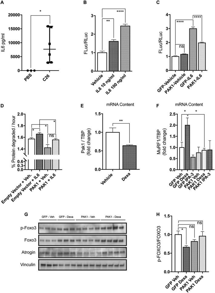
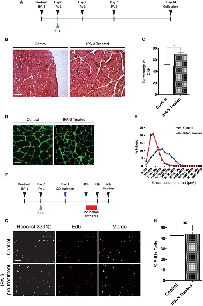
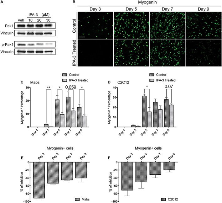
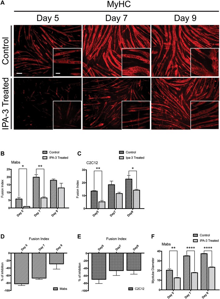
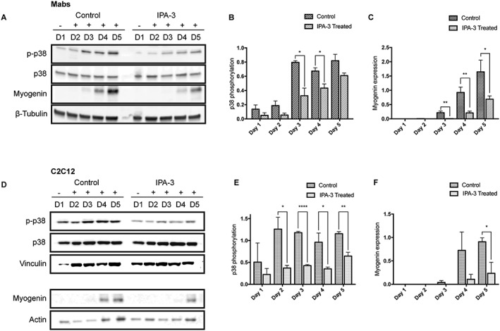
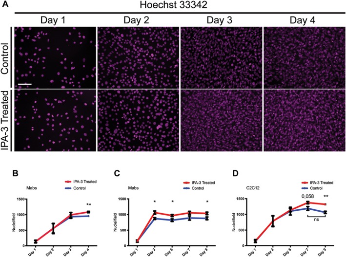
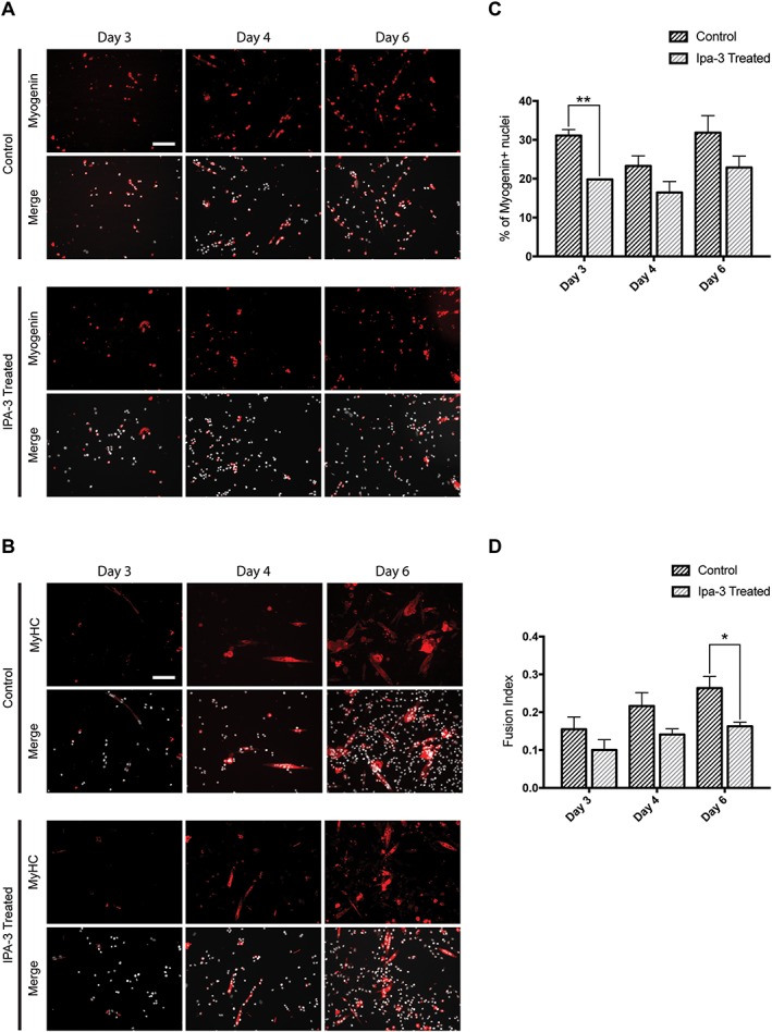
Results: We found that Pak1 expression levels are reduced during cancer-induced cachexia in the Tibialis Anterior muscles of colon adenocarcinoma C26-bearing mice and in vitro during dexamethasone-induced myotube atrophy. Electroporation of muscles of C26-bearing mice with plasmids directing the synthesis of PAK1 preserves fiber size in cachectic muscles by restraining the expression of atrogin-1 and MuRF1 and possibly by inducing myogenin expression. Consistently, the overexpression of PAK1 reduces the dexamethasone-induced expression of MuRF1 in myotubes and increases the phospho-FOXO3/FOXO3 ratio. Interestingly, the ectopic expression of PAK1 counteracts atrophy in vitro by restraining the IL6-Stat3 signalling pathway measured in luciferase-based assays and by reducing rates of protein degradation in atrophying myotubes exposed to IL6. On the other hand, we observed that the inhibition of group I Paks has no effect on myotube atrophy in vitro and is associated with impaired muscle regeneration in vivo and in vitro. In fact, we found that mice treated with the group I inhibitor IPA-3 display a delayed recovery from cardiotoxin-induced muscle injury. This is consistent with in vitro experiments showing that IPA-3 impairs myogenin expression and myotube formation in vessel-associated myogenic progenitors, C2C12 myoblasts, and satellite cells. Finally, we observed that IPA-3 reduces p38α/β phosphorylation that is required to proceed through various stages of satellite cells differentiation: activation, asymmetric division, and ultimately myotube formation.

Conclusions: Our data provide novel evidence that is consistent with group I Paks playing a central role in the regulation of muscle homeostasis, atrophy and myogenesis.

Keywords: Atrophy; Cachexia; Muscle; Paks; Regeneration.


Figures

Figure 1
Figure 1
Pak1 expression is reduced in TA muscles of cachectic C26‐bearing mice and its ectopic expression preserves myofiber area of cachectic mice by reducing the expression of atrogin‐1 and MuRF1 and possibly by inducing myogenin. (A) Representative western blot revealing total Pak1 in crude protein extracts from TA of colon adenocarcinoma‐bearing mice (C26) compared with controls (PBS). Vinculin is used as a loading control. Twenty microgram of lysates of C2C12 myoblasts previously transfected for 24 h with GFP‐PAK1 expressing plasmids have been used as controls as well as non‐transfected cells. (B) The bar graph illustrates the densitometric quantification of Pak1/vinculin signal ratio for experiments as represented in (A) (n = 4–6, unpaired t test, **P  0.01) GUSB has been used as housekeeping gene. (C) The mRNA levels of Pak1 in TA from C26‐bearing mice were determined by quantitative polymerase chain reaction (n = 6–10, unpaired t‐test, *P = 0.02). (D) Representative images of cross‐sections of TA from C26‐bearing mice or PBS‐injected ones, previously electroporated in vivo with DsRed2‐PAK1, are shown. Scale bar: 100 μm. (E) Frequency histograms showing the distribution of cross‐sectional areas of muscle fibers of TA either from PBS‐injected mice or C26‐injected ones for 14 days and transfected with empty vector or DsRed2‐PAK1. (F) The mean cross‐sectional area is shown for the four conditions described earlier (n = 219 fibers from five electroporated TA from PBS‐injected mice; n = 339 fibers from seven electroporated TA from C26‐injected mice, unpaired t‐test, ****P ≤ 0.001). Atrogin‐1 (G) and MuRF1 (H) expressions (reported in AU) inversely correlate with transfected human GFP‐PAK1 in muscle from C26‐bearing mice. On the x and y axes, the relative amount of expression of the genes indicated is reported. These numbers have been obtained by comparing the CT of the samples with that of standards of which serial dilutions have been run and probed for the same genes in the same plate. Pearson's test is shown for correlation analysis, n = 6–7. (I) The mRNA levels of myogenin in TA from C26‐bearing mice electroporated with plasmids for GFP or GFP‐PAK1 were determined by quantitative polymerase chain reaction (n = 8–9, unpaired t‐test, *P ≤ 0.05) TBP has been used as housekeeping gene. SEM is indicated in all figures. PAK1, p21 protein‐activated kinase 1; TA, tibialis anterior; AU, arbitrary units; GUSB, β‐glucuronidase; TBP, Tata‐Binding Protein.
Figure 2
Figure 2
PAK1 overexpression exerts anti‐atrophic effects in two models of in vitro C2C12 atrophy. (A) The circulating levels of IL6 in the plasma of C26‐bearing mice is drastically increased with respect to PBS‐injected mice (n = 3–6, Mann Whitney's test, *P = 0.02). (B) Upon 5 h treatment with 10 or 100 ng/mL murine IL6, C2C12 myoblasts transiently expressing Stat3 4X‐FLuc reporter plasmids induce FLuc (n = 3, one‐way anova followed by Dunnett's test, **P ≤ 0.01, ****P ≤ 0.0001). (C) IL6‐induced Stat3‐FLuc signal is reduced in myoblasts expressing GFP‐PAK1 with respect to those expressing GFP. Myoblasts treated for 5 h with IL6 (100 ng/mL) were previously transfected with Stat3 4X‐FLuc plasmid, Renilla luciferase plasmids, and GFP or PAK1‐expressing plasmids. The results of three independent experiments are shown. Mean is reported (n = 14, unpaired t‐test, ***P = 0.0002). (D) Rates of long‐lived protein degradation were measured in myotubes transfected on the third day of differentiation with plasmids for pDSRed2‐PAK1 or empty vector and differentiated for one more day, when they were exposed for 24 h to 10 ng/mL IL6 (n = 4, unpaired t‐test, *P ≤ 0.05). (E) The mRNA content of Pak1 is reduced in atrophying myotubes exposed for 24 h to 10 μM dexamethasone (n = 6, Mann Whitney's test, **P = 0.002, ****P ≤ 0.0001). (F) Myotubes transfected on the third day of differentiation with plasmids for GFP‐PAK1 or only GFP were exposed for 24 h on the fourth day of differentiation to vehicle or 1 μM dexamethasone or 10 μM IPA‐3. In these conditions, the mRNA levels of MuRF1 were determined by quantitative polymerase chain reaction (n = 4, unpaired t‐test, *P ≤ 0.05). (G) Myoblasts were transfected, as indicated earlier, for 24 h, total protein was then extracted. Immunoblot analysis reveals the protein content of p‐FOXO3, FOXO3, atrogin‐1, and vinculin that is used as loading control. (H) Quantification of the ratio between p‐FOXO3 over total FOXO3 is shown (n = 6–8, unpaired t‐test, *P ≤ 0.05). SEM is indicated in all figures. IL6, interleukin‐6; FLuc, firefly luciferase; PAK1, p21 protein‐activated kinase 1; TBP, Tata‐Binding Protein.
Figure 3
Figure 3
IPA‐3 in vivo treatment delays muscle regeneration. (A) Graphical representation of the IPA‐3 treatments prior and following CTX treatment. (B) Representative images of haematoxylin and eosin histological staining on mouse TA sections 14 days after CTX injection. Scale bar: 100 μm. (C) The bar graph shows the percentage of CNF 14 days after CTX injury in the TA of control and IPA‐3 treated mice (n = 2, unpaired t‐test, *P ≤ 0.05). (D) Immunofluorescence images showing laminin expression in mouse TA sections 14 days after CTX injection. Scale bar: 50 μm. (E) Graphical representation of CSA distribution in TA of IPA‐3 treated and control mice 14 days after CTX injury. The values represent the mean of two muscle samples (n = 2). (F) Schematic representation of the EdU labelling of SC following in vivo IPA‐3 pre‐treatment and CTX injury. (G) Immunofluorescence microphotographs of SCs 24 h upon EdU pulse (green). Scale bar: 100 μm. (H) Bar plot showing the percentage of EdU positive SCs for the experiment reported in G (n = 3, unpaired t‐test). SEM is indicated in all figures. CTX, cardiotoxin; CNF, centrally nucleated fiber; TA, tibialis anterior.
Figure 4
Figure 4
IPA‐3 treatment reduces myogenin expression throughout Mabs and C2C12 myogenic differentiation process. (A) Immunoblot analysis revealing p‐Pak1 (S144) and total Pak1 in C2C12 protein extracts following 2 h treatment with various concentrations of the inhibitor IPA‐3 or vehicle (dimethyl sulfoxide). Pak1 and p‐Pak1 signals have been developed on separate filters and vinculin serves as a loading control. (B) Immunofluorescence microphotographs of Mabs labelled with an antibody against myogenin (green). (C, D) Bar plot representing the percentage of myogenin positive cells for Mabs and C2C12, respectively. (E) The graph illustrates the percentage difference of myogenin positive cells during Mabs myogenic differentiation program. (F) Graph showing the different percentage of myogenin positive cells during C2C12 differentiation. The values represented are means of at least three independent experiments + SEM. Statistical significance has been evaluated using the unpaired t‐test, *P ≤ 0.05, **P ≤ 0.01. Scale bar: 100 μm. PAK1, p21 protein‐activated kinase 1; Mabs, mesoangioblasts.
Figure 5
Figure 5
IPA‐3 treatment affects MyHC expression and myotube formation of differentiating myogenic cells. (A) Immunofluorescence images, showing MyHC expression (red) in IPA‐3 treated differentiating Mabs. The insets display a higher magnification (×20). (B, C) Average fusion index for Mabs and C2C12 following IPA‐3 treatment. (D, E) Bar plots representing the percentage of inhibition for the fusion index between IPA‐3 treated and control Mabs or C2C12. (F) Bar plot of Mabs‐derived myotube diameter (μm). The plotted values are means of three independent experiments + SEM. Statistical significance has been evaluated using the unpaired t‐test, *P ≤ 0.05, **P ≤ 0.01, ****P ≤ 0.0001. Scale bar for ×20 magnification: 50 μm. Scale bar for ×10 magnification: 100 μm. MyHC, myosin heavy chain; Mabs, mesoangioblasts.
Figure 6
Figure 6
IPA‐3 treatment reduces p38 phosphorylation during Mabs and C2C12 differentiation. Representative western blot revealing total p38, p‐p38, and myogenin expression in crude protein extracts from IPA‐3 treated or control Mabs (A) and C2C12 (D) during 5 days of myogenic differentiation. β‐Tubulin, vinculin, or actin are used as loading controls. The bar graphs illustrate the ratio between p‐p38 and total p38 signals during Mabs (B) or C2C12 (E) differentiation, as determined by the densitometric quantitation of western blots such as the one in panel A and D. The bar graphs represent the densitometric quantitation of myogenin expression for Mabs (C) and C2C12 (F). The values are mean of at least three independent experiments + SEM. Statistical significance was evaluated by the unpaired t‐test, *P ≤ 0.05, **P ≤ 0.01, ****P ≤ 0.0001. Mabs, mesoangioblasts.
Figure 7
Figure 7
IPA‐3 treatment delays Mabs cell cycle exit without affecting proliferation rate. (A) The panels show Mabs nuclei stained with Hoechst 33342 (magenta) at four time points after plating in growth medium supplemented with DMSO (control) or with IPA‐3. (B) The graph illustrates the number of Mabs nuclei/field during the first 4 days of differentiation. (C, D) The graphs show the number of nuclei/field during 9 days of differentiation for Mabs and C2C12, respectively. Field area is approx. 2,84 * 105 μm2. The values are mean of at least three independent experiments + SEM. Statistical significance has been evaluated using the unpaired t‐test, *P ≤ 0.05, **P ≤ 0.01. Scale bar: 100 μm. Mabs, mesoangioblasts.
Figure 8
Figure 8
IPA‐3 treatment affects satellite cells differentiation. (A, B) Immunofluorescence showing myogenin (red) and myosin heavy chain (MyHC; red) expression during SC differentiation. Nuclei are represented in grey. (C) Bar plot of the percentage of myogenin positive cells. (D) The bar graph represents fusion index during SC differentiation. The values are mean of three independent experiments + SEM. Statistical significance has been evaluated using the unpaired t‐test, *P ≤ 0.05, **P ≤ 0.01. Scale bar: 100 μm.

References

    1. Piccirillo R, Demontis F, Goldberg AL, Perrimon N. Mechanisms of muscle growth and atrophy in mammals and Drosophila. Dev Dyn 2014;243:201–215. - PMC - PubMed
    1. Yin H, Price F, Rudnicki MA. Satellite cells and the muscle stem cell niche. Physiol Rev 2013;93:23–67. - PMC - PubMed
    1. He WA, Berardi E, Cardillo VM, Acharyya S, Aulino P, Thomas‐Ahner J, et al. NF‐κB‐mediated Pax7 dysregulation in the muscle microenvironment promotes cancer cachexia. J. Clin. Invest. American Society for Clin Investig 2013;123:4821–4835. - PMC - PubMed
    1. Wigmore SJ, Plester CE, Ross JA, Fearon KC. Contribution of anorexia and hypermetabolism to weight loss in anicteric patients with pancreatic cancer. Br J Surg 1997;84:196–197. - PubMed
    1. Fearon K, Strasser F, Anker SD, Bosaeus I, Bruera E, Fainsinger RL, et al. Definition and classification of cancer cachexia: an international consensus. Lancet Oncol Elsevier 2011;12:489–495. - PubMed

Publication types

MeSH terms