

A Case of Corneal Neovascularization Misdiagnosed as Total Limbal Stem Cell Deficiency
- PMID: 29781927
- PMCID: PMC6037563
- DOI: 10.1097/ICO.0000000000001631
A Case of Corneal Neovascularization Misdiagnosed as Total Limbal Stem Cell Deficiency
Abstract
Purpose: To report a case of corneal neovascularization misdiagnosed as total limbal stem cell deficiency (LSCD).
Methods: This is a case report of a 61-year-old woman who has a history of bilateral idiopathic scleritis, keratitis, and uveitis for more than 20 years. She was diagnosed with total LSCD in her left eye based on clinical presentation alone and was confirmed as a candidate for limbal transplantation at several major tertiary eye care centers in the United States. After referral to the Stein Eye Institute, in vivo confocal microscopy (IVCM) and anterior segment optical coherence tomography (AS-OCT) were performed to clarify the diagnosis.
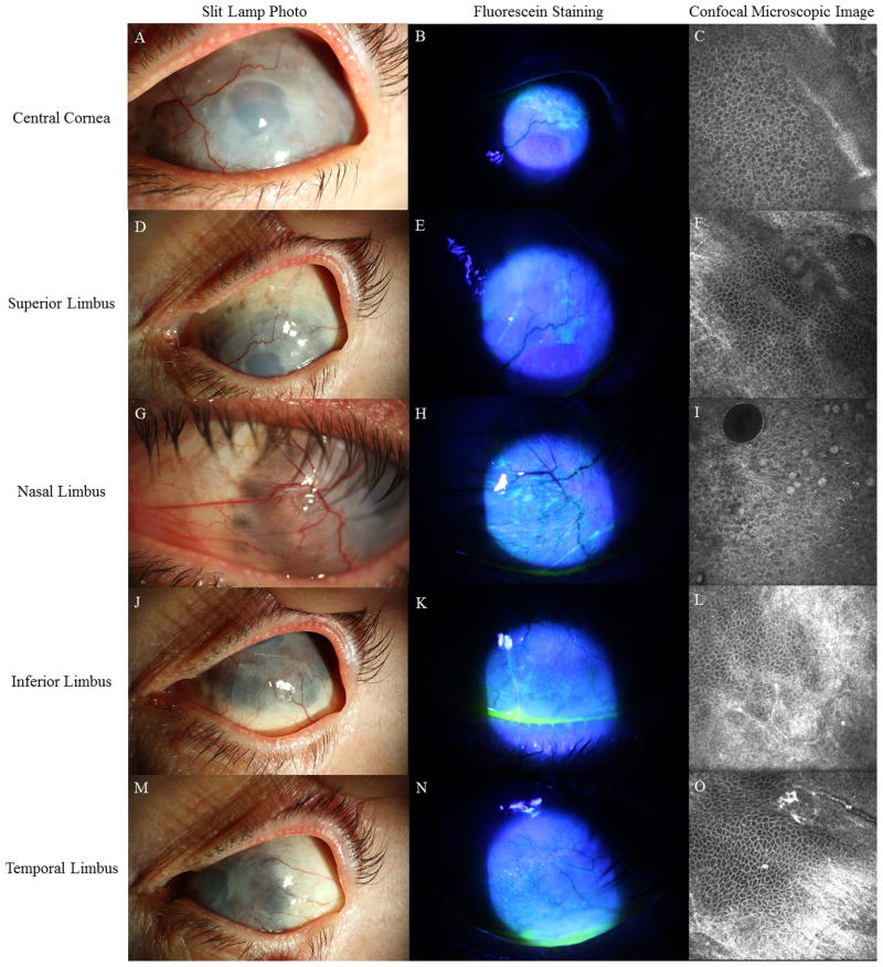
Results: Slit-lamp examination of the left eye revealed 360-degree severe thinning at the limbus and peripheral corneal pannus and neovascularization that spared the central cornea, a smooth epithelium without fluorescein staining at the central cornea, an uneven surface, and pooling of fluorescein at the peripheral cornea accompanied by minimal fluorescein staining of the sectoral peripheral epithelium. IVCM showed that epithelial cells in the central cornea exhibited a corneal phenotype and that the morphology of the epithelium in all limbal regions except the nasal limbus was normal. Epithelial cellular density and thickness were within the normal range. AS-OCT showed severe thinning in the limbus and a normal epithelial layer in the cornea and limbus. Based on the findings of IVCM and AS-OCT, we concluded that the patient had minimal LSCD, and limbal stem cell transplantation was not recommended.
Conclusions: Clinical presentation alone is insufficient to correctly diagnose LSCD in complex cases. Additional diagnostic tests, such as IVCM, are necessary to confirm the diagnosis before any surgical intervention.
Conflict of interest statement
Conflict of interest: None of the authors has financial interests related to the content of the manuscripts.
Figures

References
-
- Miri A, Al-Aqaba M, Otri AM, et al. In vivo confocal microscopic features of normal limbus. Br J Ophthalmol. 2012;96:530–536. - PubMed
-
- Yang Y, Hong J, Deng SX, et al. Age-related changes in human corneal epithelial thickness measured with anterior segment optical coherence tomography. Invest Ophthalmol Vis Sci. 2014;55:5032–5038. - PubMed
Publication types
MeSH terms
Grants and funding
LinkOut - more resources
Full Text Sources
Other Literature Sources
Medical
Research Materials
