Review
. 2018 Jun;93(6):592-596.
doi: 10.1002/cyto.a.23488. Epub 2018 May 21.

OMIP-047: High-Dimensional phenotypic characterization of B cells

Affiliations
Review

OMIP-047: High-Dimensional phenotypic characterization of B cells

Thomas Liechti et al. Cytometry A. 2018 Jun.
No abstract available


Figures

Figure 1
Figure 1
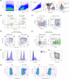
Characterization of B cell subsets by flow cytometry. (A) A Time versus SSC‐A gate was set to exclude fluorescence intensity fluctuations due to eventual irregular acquisition of the flow cytometer. Single cells were further defined based on SSC‐A and FSC‐A and a gate for doublet discrimination based on FSC‐H/FSC‐A. B cells were defined as Dump‐negative (Dump set as CD3, CD14, CD16 positive and dead cells) and CD19‐positive cells. A major B cell subset discrimination is based on CD10 and, therefore, B cells were divided into CD10 and CD10+ subpopulations. (B) CD10+ B cells can be further refined into transitional B cells (CD38+IgD+). The gating of CD38+ cells was done based on a FMO control for CD38 AF700 shown in the left panel. (C) CD10 B cells comprise IgD+ unswitched B cells (CD38IgD+), memory B cells (CD38IgD) and plasmablasts (PB; CD38highIgD), which can be defined based on the surface expression pattern of CD38 and IgD. The unswitched IgD+ fraction can be further divided in naïve (CD27IgDhigh) and marginal zone (MZ) B cells (CD27+IgD+) as shown in the right panel. (D) Memory B cells derived from the memory B cell gate (CD38IgD) in Figure 1C can be further divided in subsets based on their maturation state by expression patterns of CD21 and CD27 as described 3, 18, 29. In HIV‐1 infected patients the distribution is known to be skewed toward activated (AM) and tissue‐like (TLM) memory B cells 3, 18, 29. The analysis of cell samples from HIV‐1 infection (Figure 1D, right panel) was thus useful to verify sufficient resolution of CD21 staining. (E) Alternatively, memory B cells can be divided in subsets based on their expressed B cell receptor isotype. The panel includes antibodies specific for IgG1, IgG3, and IgA. IgA‐expressing switched memory B cells (right panel) are defined from cells within the “Non‐IgG1 and ‐IgG3” gate (left panel, transparent green gate). (F) To characterize the potential migratory activity of B cell subsets, we included antibodies for the chemokine receptors CCR7, CXCR3, CXCR4 and CXCR5 in our panel. To measure responsiveness to IL‐21, an important cytokine for development of high‐affinity antibody responses, IL‐21R levels were measured. The resolution of anti‐CXCR4, ‐CXCR5, and –IL‐21R antibodies was sufficient as judged by staining differences between the full stained sample (blue) and the corresponding FMO control (grey). (G) Resolution of CCR7 proved high so that FMO was not necessary to define positive populations. (H) In healthy donors the frequency of CXCR3‐ and Ki67‐expressing B cells is genuinely low necessitating FMO controls. FMO control and fully stained sample are shown as a dot plot in combination with CD27. Both markers show sufficient separation in combination with full staining panel. Data in panels 1A, B, C, and E are from measurements of the same sampling time point of one healthy donor. Data shown in right panel of Figure 1D is from measurements of PBMC from a chronically HIV‐1 infected individual. All percentages are related to total DumpCD19+ B cells except in Figures 1D and E where percentages are related to memory B cells as gated in Figure 1C. Percentages stated in brackets in Figure 1B are related to total CD10+ B cells. [Color figure can be viewed at wileyonlinelibrary.com]

References

    1. Malaspina A, Moir S, Ho J, Wang W, Howell ML, O'Shea MA, Roby GA, Rehm CA, Mican JM, Chun TW, et al. Appearance of immature/transitional B cells in HIV‐infected individuals with advanced disease: Correlation with increased IL‐7. Proc Natl Acad Sci U S A 2006;103:2262–2267. - PMC - PubMed
    1. Sugalski JM, Rodriguez B, Moir S, Anthony DD. Peripheral blood B cell subset skewing is associated with altered cell cycling and intrinsic resistance to apoptosis and reflects a state of immune activation in chronic hepatitis C virus infection. J Immunol 2010;185:3019–3027. - PMC - PubMed
    1. Moir S, Fauci AS. Insights into B cells and HIV‐specific B‐cell responses in HIV‐infected individuals. Immunol Rev 2013;254:207–224. - PubMed
    1. Doi H, Tanoue S, Kaplan DE. Peripheral CD27‐CD21‐ B‐cells represent an exhausted lymphocyte population in hepatitis C cirrhosis. Clin Immunol 2014;150:184–191. - PMC - PubMed
    1. Moir S, Buckner CM, Ho J, Wang W, Chen J, Waldner AJ, Posada JG, Kardava L, O'Shea MA, Kottilil S. B cells in early and chronic HIV infection: evidence for preservation of immune function associated with early initiation of antiretroviral therapy. Blood 2010;116:5571–5579. - PMC - PubMed

MeSH terms

Substances