

Magnolol and Honokiol Attenuate Apoptosis of Enterotoxigenic Escherichia Coli-Induced Intestinal Epithelium by Maintaining Secretion and Absorption Homeostasis and Protecting Mucosal Integrity
- PMID: 29782483
- PMCID: PMC5990993
- DOI: 10.12659/MSM.910350
Magnolol and Honokiol Attenuate Apoptosis of Enterotoxigenic Escherichia Coli-Induced Intestinal Epithelium by Maintaining Secretion and Absorption Homeostasis and Protecting Mucosal Integrity
Abstract
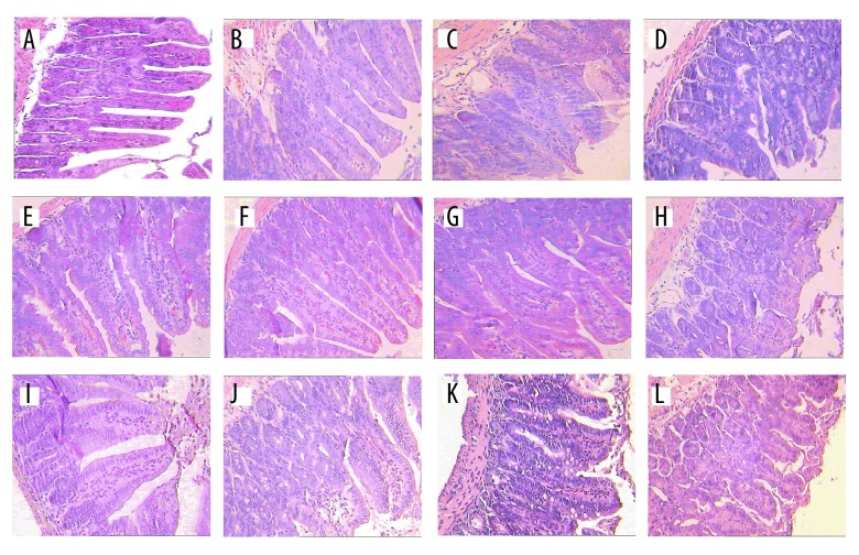
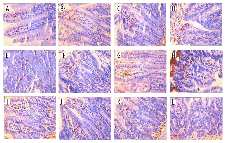
BACKGROUND The cortex of Magnolia officinalis has long been used as an element of traditional Chinese medicine for the treatment of anxiety, chronic bronchitis, and gastrointestinal dysfunction. This study aimed to elucidate the underlying mechanism of its functional ingredients (magnolol and honokiol) in modifying the secretion and absorption homeostasis and protecting mucosal integrity in an Enterotoxigenic Escherichia coli (ETEC)-induced diarrhea mouse model. MATERIAL AND METHODS This study established a diarrhea mouse model infected by ETEC at a dosage of 0.02 ml/g live body weight (BW) in vivo. Magnolol or honokiol was followed by an intraperitoneal administration at dosages of 100, 300, and 500 mg/kg BW according to a 3×3 factorial arrangement. The useful biomarkers for evaluating the integrity of intestinal tract and histologic injury were analyzed and morphological development (including villus height, crypt depth, and ratio of villus height to crypt depth) and the expressions of inflammatory cytokines were determined by real-time PCR. RESULTS The results showed that magnolol and honokiol (500 mg/kg BW) reduced the concentrations of NO, DAO, and DLA, and iNOS activity, and the mRNA expressions of the interferon gamma (IFN-γ) and interleukin 10 (IL-10), and inhibited intestinal epithelial cell apoptosis. Magnolol and honokiol (300 mg/kg BW) elongated the villus height and crypt depth and decreased the number of goblet cells and the ratio of villus height to crypt depth. CONCLUSIONS The current results indicate that magnolol and honokiol enhance the intestinal anti-inflammatory capacities, elongate the villus height and crypt depth, and reduce goblet cell numbers to inhibit the intestinal epithelium apoptosis and effectively protect the intestinal mucosa. These results show that magnolol and honokiol protect the intestinal mucosal integrity and regulate gastrointestinal dysfunction.
Conflict of interest statement
None.
Figures
References
-
- Lucas ML. Diarrhoeal disease through enterocyte secretion: A doctrine untroubled by proof. Exp Physiol. 2010;95(4):479–84. - PubMed
MeSH terms
Substances
Grants and funding
LinkOut - more resources
Full Text Sources
Other Literature Sources
