. 2018 Sep;155(3):865-879.e12.
doi: 10.1053/j.gastro.2018.05.027. Epub 2018 May 18.

Impaired TFEB-Mediated Lysosome Biogenesis and Autophagy Promote Chronic Ethanol-Induced Liver Injury and Steatosis in Mice

Affiliations

Impaired TFEB-Mediated Lysosome Biogenesis and Autophagy Promote Chronic Ethanol-Induced Liver Injury and Steatosis in Mice

Xiaojuan Chao et al. Gastroenterology. 2018 Sep.

Abstract

Background & aims: Defects in lysosome function and autophagy contribute to the pathogenesis of alcoholic liver disease. We investigated the mechanisms by which alcohol consumption affects these processes by evaluating the functions of transcription factor EB (TFEB), which regulates lysosomal biogenesis.

Methods: We performed studies with GFP-LC3 mice, mice with liver-specific deletion of TFEB, mice with disruption of the transcription factor E3 gene (TFE3-knockout mice), mice with disruption of the Tefb and Tfe3 genes (TFEB and TFE3 double-knockout mice), and Tfebflox/flox albumin cre-negative mice (controls). TFEB was overexpressed from adenoviral vectors or knocked down with small interfering RNAs in mouse livers. Mice were placed on diets of regular ethanol feeding plus an acute binge to induce liver damage (ethanol diet); some mice also were given injections of torin-1, an inhibitor of the kinase activity of the mechanistic target of rapamycin (mTOR). Liver tissues were collected and analyzed by immunohistochemistry, immunoblots, and quantitative real-time polymerase chain reaction to monitor lysosome biogenesis. We analyzed levels of TFEB in liver tissues from patients with alcoholic hepatitis and from healthy donors (controls) by immunohistochemistry.

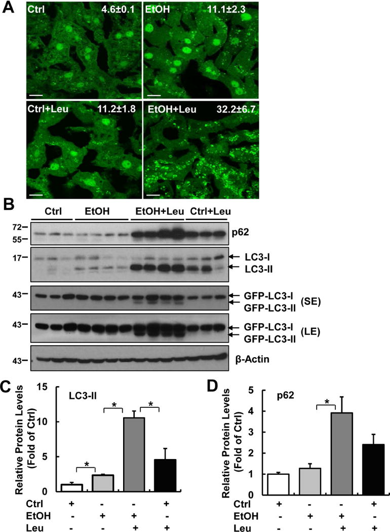
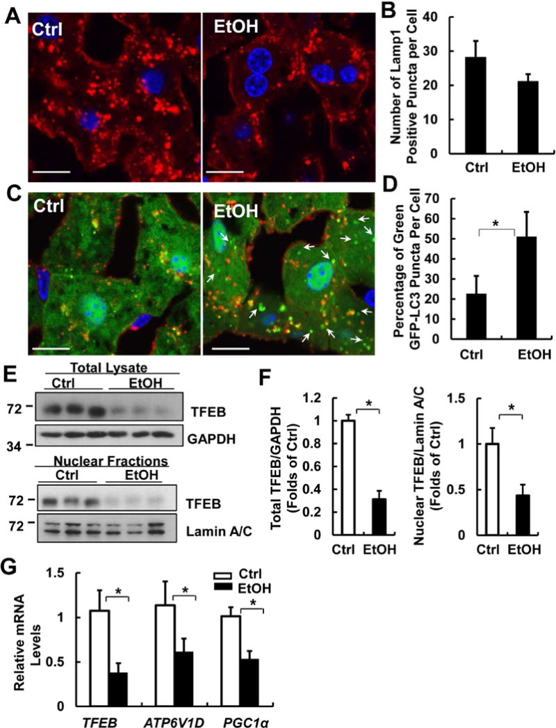
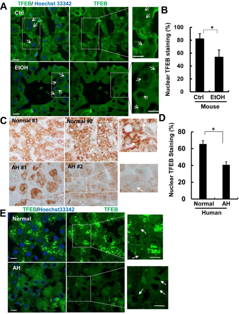
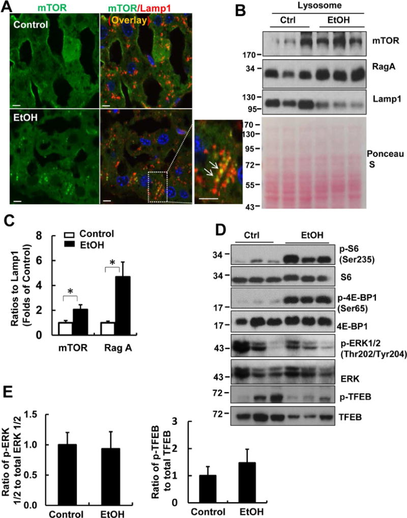
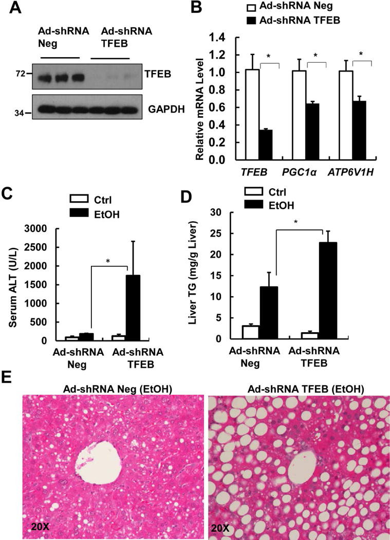
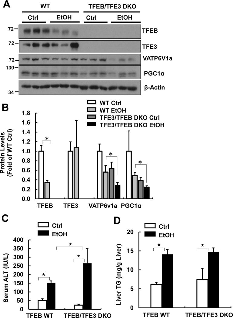
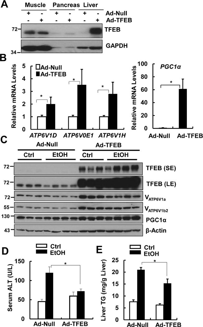
Results: Liver tissues from mice on the ethanol diet had lower levels of total and nuclear TFEB compared with control mice, and hepatocytes had decreased lysosome biogenesis and autophagy. Hepatocytes from mice on the ethanol diet had increased translocation of mTOR into lysosomes, resulting in increased mTOR activation. Administration of torin-1 increased liver levels of TFEB and decreased steatosis and liver injury induced by ethanol. Mice that overexpressed TFEB in the liver developed less severe ethanol-induced liver injury and had increased lysosomal biogenesis and mitochondrial bioenergetics compared with mice carrying a control vector. Mice with knockdown of TFEB and TFEB-TFE3 double-knockout mice developed more severe liver injury in response to the ethanol diet than control mice. Liver tissues from patients with alcohol-induced hepatitis had lower nuclear levels of TFEB than control tissues.

Conclusions: We found that ethanol feeding plus an acute binge decreased hepatic expression of TFEB, which is required for lysosomal biogenesis and autophagy. Strategies to block mTOR activity or increase levels of TFEB might be developed to protect the liver from ethanol-induced damage.

Keywords: Fatty Liver; Gene Regulation; Hepatic Protection; Mouse Model.


Conflict of interest statement

Conflict of Interest: These authors declare that they have nothing to disclose.

Figures

Figure 1
Figure 1. Gao-binge induces autophagic flux in mouse livers
Male GFP-LC3 transgenic mice were treated with Gao-binge model. One dose of Leupeptin (40 mg/kg, i.p) was given to mice prior to the gavage. Mice were sacrificed 8 hours later after the gavage. (A) Representative confocal microscopy images from liver cryosections are shown. GFP-LC3 puncta per cell in each group were quantified (n=3-4). Ctrl: control diet+maltose gavage; EtOH: ethanol diet+ ethanol gavage. Data are means ± SE. More than 60 cells were counted in each mouse. Scale Bar: 10 μm. (B) Total liver lysates were subjected to western blot analysis. SE: short exposure; LE: long exposure. (C & D) Densitometry analysis of (B). The levels of p62 and LC3-II were normalized to the loading control (β-Actin). Data represent means ± SE (n= 3-4). *p<0.05; one-way ANOVA analysis.
Figure 2
Figure 2. Gao-binge decreases the number of lysosomes with increased accumulation of autophagosomes and inhibits TFEB in mouse livers
Male GFP-LC3 transgenic mice were treated with Gao-binge model and cryosections of mouse livers were subjected to immunostaining for Lamp1 followed by confocal microscopy. Representative images of immunostaining of Lamp1 (A) and quantified number of Lamp1 positive vesicles are shown (B). Scale Bar: 10 μm. Data are means ± SE (n=3-4). More than 50 cells were counted in each mouse. (C) Representative images of the colocalization of Lamp1 with GFP-LC3 puncta are shown. Arrows denote the green-only GFP-LC3 puncta. (D) Percentage of GFP-LC3 puncta that are not colocalized with Lamp1 positive vesicles. Data are means ± SE (n=3-4). More than 50 cells were counted in each mouse. *p<0.05; Student t test. (E) Male C57BL/6J WT mice were treated with Gao-binge model. Total lysates and nuclear fractions from mouse livers were subjected to western blot analysis. (F) Densitometry analysis of (E). (G) mRNA from mouse livers was used for qPCR. Results were normalized to 18s and expressed as fold change compared to Ctrl group. Data shown are means ± SE (n =4). *p<0.05; Student t test.
Figure 3
Figure 3. Decreased nuclear TFEB proteins in Gao-binge alcohol mouse livers and human alcoholic hepatitis
Male C57BL/6J WT mice were treated with Gao-binge model. Cryosections of mouse livers were subjected to immunostaining for TFEB and nuclei were stained with Hoechst33342 followed by confocal microscopy. Representative images are shown (A). Arrows denote the nuclear staining of TFEB. Right panels are enlarged photographs from the boxed areas. Scale Bar: 10 μm. (B) The percentage of hepatocytes with nuclear TFEB staining were quantified from 5 fields of 40X magnification from 3 mice of each group. Data are means ± SE. *p<0.05; Student t test. (C) Immunohistochemical staining of liver TFEB of alcoholic hepatitis (AH) patients or healthy donors (normal liver), and representative images are shown. (D) The percentage of hepatocytes with nuclear TFEB staining were quantified from 4 fields of 40X magnification (n=6 healthy controls; and n=6 AH). Data are means ± SE. *p<0.05; Student t test. (E) Cryo-liver sections of AH patient and healthy donor were subjected immunostaining of TFEB and nuclear were stained with Hoechst33342 followed by confocal microscopy. Images in right panels were enlarged photographs from the boxed areas in the left panels. Arrows denote the nuclear TFEB staining.
Figure 4
Figure 4. Gao-binge activates mTOR in mouse livers
Male C57BL/6J WT mice were treated with Gao-binge model. (A) Cryosections of mouse livers were subjected to immunostaining for mTOR (green) and Lamp1 (red) followed by confocal microscopy. Representative images are shown. Right panel is an enlarged photograph from the boxed area. Arrows denote the colocalization of mTOR with Lamp1. Scale Bar: 10 μm. (B) Lysosomal fractions from mouse livers were subjected to western blot analysis. Membrane was pre-stained with Ponceau S as a loading control. (C) Densitometry analysis of (B) are shown (means ± SE, n=3). *p<0.05; Student t test. (D) Total liver lysates were subjected to western blot analysis. (E) Densitometry analysis of (D) are shown (means ± SE, n=3).
Figure 5
Figure 5. Knockdown of hepatic TFEB exacerbates Gao-binge-induced liver injury and steatosis
Male C57BL/6J WT mice were injected with Ad-shRNA negative and Ad-shRNA TFEB (1×10⁹ PFU/mouse via tail vein) followed by Gao-binge model. (A) Total liver lysates were subjected to western blot analysis. (B) Hepatic mRNA was extracted followed by qPCR (n=4-6). Data are means ± SE. *p<0.05; Student t test. (C) Serum ALT levels and (D) hepatic TG levels were quantified. Data are means ± SE (n=4-6). * p<0.05; One-way ANOVA analysis. (E) Representative images of H& E staining are shown.
Figure 6
Figure 6. TFEB/TFE3 DKO mice are more susceptible to Gao-binge-induced liver injury
Male TFEB/TFE3 DKO and matched WT mice were treated with Gao-binge model. (A) Total liver lysates were subjected to western blot analysis and (B) densitometry analysis of (A). Serum ALT (C) and liver TG levels (D) were measured. Data are means ± SE (n=3-10). *p<0.05; One-way ANOVA analysis.
Figure 7
Figure 7. Overexpression of TFEB protects against alcohol-induced steatosis and liver injury
Male WT C57BL/6J mice were injected with Ad-Null and Ad-TFEB (5×10⁸ PFU/mouse via tail vein) followed by Gao-binge model. (A) Total muscle, pancreas and liver tissue lysates from mice that were treated with control diet+maltose gavage were subjected to western blot analysis. (B) Hepatic mRNA was extracted followed by qPCR (n=4-6). Data are means ± SE (n=4-6). * p<0.05; Student t test. (C) Total liver lysates were subjected to western blot analysis. SE: short exposure; LE: long exposure. (D) Serum ALT levels and (E) hepatic TG levels were quantified. Data are means ± SE (n=4-6). * p<0.05; one-way ANOVA analysis.

Comment in

References

    1. Liangpunsakul S, Haber P, McCaughan GW. Alcoholic Liver Disease in Asia, Europe, and North America. Gastroenterology. 2016;150:1786–97. - PMC - PubMed
    1. Nagy LE, Ding WX, Cresci G, et al. Linking Pathogenic Mechanisms of Alcoholic Liver Disease With Clinical Phenotypes. Gastroenterology. 2016;150:1756–68. - PMC - PubMed
    1. Gao B, Bataller R. Alcoholic liver disease: pathogenesis and new therapeutic targets. Gastroenterology. 2011;141:1572–85. - PMC - PubMed
    1. Williams JA, Manley S, Ding WX. New advances in molecular mechanisms and emerging therapeutic targets in alcoholic liver diseases. World J Gastroenterol. 2014;20:12908–33. - PMC - PubMed
    1. Ding WX, Li M, Chen X, et al. Autophagy reduces acute ethanol-induced hepatotoxicity and steatosis in mice. Gastroenterology. 2010;139:1740–52. - PMC - PubMed

Publication types

Substances