Review
. 2018 May 20;8(5):349.
doi: 10.3390/nano8050349.

Graphene-Based Nanomaterials for Tissue Engineering in the Dental Field

Affiliations
Review

Graphene-Based Nanomaterials for Tissue Engineering in the Dental Field

Riccardo Guazzo et al. Nanomaterials (Basel). .

Abstract

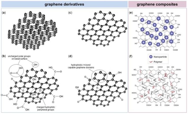
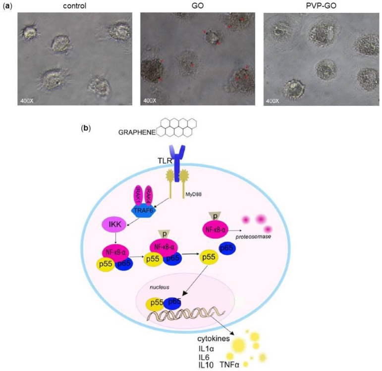
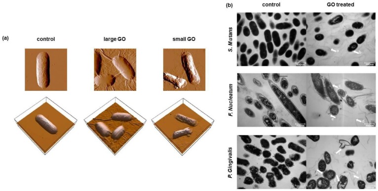
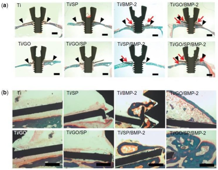
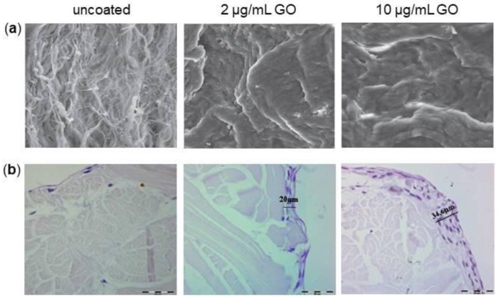
The world of dentistry is approaching graphene-based nanomaterials as substitutes for tissue engineering. Apart from its exceptional mechanical strength, electrical conductivity and thermal stability, graphene and its derivatives can be functionalized with several bioactive molecules. They can also be incorporated into different scaffolds used in regenerative dentistry, generating nanocomposites with improved characteristics. This review presents the state of the art of graphene-based nanomaterial applications in the dental field. We first discuss the interactions between cells and graphene, summarizing the available in vitro and in vivo studies concerning graphene biocompatibility and cytotoxicity. We then highlight the role of graphene-based nanomaterials in stem cell control, in terms of adhesion, proliferation and differentiation. Particular attention will be given to stem cells of dental origin, such as those isolated from dental pulp, periodontal ligament or dental follicle. The review then discusses the interactions between graphene-based nanomaterials with cells of the immune system; we also focus on the antibacterial activity of graphene nanomaterials. In the last section, we offer our perspectives on the various opportunities facing the use of graphene and its derivatives in associations with titanium dental implants, membranes for bone regeneration, resins, cements and adhesives as well as for tooth-whitening procedures.

Keywords: antibacterial activity; bone regeneration; dental implant; dental stem cells; graphene; nanomaterials.


Conflict of interest statement

The authors declare no conflict of interest. No grants were awarded in relation to this study.

Figures

Figure 1
Figure 1
Schematic representation of different graphene-based nanomaterials. (a) Few-Layered Graphene (FLG), (b) Graphene Oxide (GO), (c) graphene nanosheets and (d) reduced Graphene Oxide (rGO) belong to the graphene derivatives group; (e) GO nanoparticle composite and (f) GO polymer composite are composites of graphene. Reproduced with permissions from [25,26].
Figure 2
Figure 2
Morphological observations of cells after interactions with graphene-based nanomaterials. (a) Transmission Electron Microscopy (TEM) images of Mouse Embryo Fibroblasts (MEFs) treated with GO-high (GO-h), GO-medium (GO-m) and GO-low (GO-l) at 50 µg/mL for 24 h. On bottom, high-magnification images of the boxed-in photos on top are represented. The white and black arrows indicate GO aggregates inside and outside cells, respectively. (b) Scanning Electron Microscopy (SEM) images of cell membrane damage incurred by A549 cells as a result of GO nanosheets exposure observed during different phases of incubation. On bottom, high-magnification images of the boxed-in photos on top are represented. Reproduced with permissions from [40,42].
Figure 3
Figure 3
Effect of graphene-based nanomaterials on the osteogenic differentiation of Mesenchymal Stem Cells (MSCs). (a) Evaluation of matrix mineralization by means of Alizarin Red S (ARS) staining (top panel) and quantification (bottom panel). MSCs were grown for 21 days in osteogenic differentiation medium on GO nanosheets. The level of mineralization in the 0.1 μg/mL GO group was significantly higher than the other two conditions. (b) Human BM-MSCs cultured on 3D graphene foams for 4 days show protrusions up to 100 μm in length (yellow arrowheads) that extended from the cell bodies (black arrows), as evidenced by SEM images (top panel). These 3D substrates were also found to promote the expression of the osteogenic markers Osteocalcin (OCN) and Osteopontin (OPN), as displayed by immunofluorescence images (bottom panel). (c) SEM images of the rGO-coated Hap nanocomposites showing that Hydroxyapatite (HAp) particles were partly covered and interconnected by an network of rGO nanosheets (top panel). ARS staining performed at 21 days reveals that rGO-coated HAp nanocomposites significantly increase calcium deposits in MC3T3-E1 cells compared to the non-treated control and rGO or HAp alone (bottom panel). (d) Schematic representation of the study: human BM-MSCs were seeded onto rGO substrates, then exposed to Pulsed Electromagnetic Fields (PEMFs) (top panel). The rGO+PEMFs group exhibited the strongest staining as evidenced by ARS staining performed after 2 weeks from cells seeding. Reproduced with permissions from [50,51,53,62].
Figure 4
Figure 4
Interactions of graphene-based nanomaterials with Dental Stem Cells (DSCs). (a) SEM images showing that Dental Pulp Stem Cells (DPSCs) can efficiently adhere and proliferate on GO substrates for 3 and 5 days (top panel). DPSCs on GO present higher expression compared to glass (control) for all genes tested both at 7 and 14 days (bottom panel). (b) SEM images of films composed of Silk Fibroin (SF), GO and GO-SF mixture (3:1) at different magnifications (top panel). Immunofluorescence staining of the actin cytoskeleton showing a higher adhesion of Periodontal Ligament Stem Cells (PDLSCs) on GO and on the GO-SF composite film rather than on SF alone at 7 days. (c) TEM images of GO, Thermally Reduced Graphene Oxide (TRGO) and Nitrogen-doped graphene (N-Gr) (top panel). Confocal microscopy images of human Dental Follicle Progenitor Cells (DFPCs) seeded on GO, TRGO and N-Gr at 40 µg/mL showing staining of cytoskeleton actin filaments (green) and nuclei (red) (bottom panel). Reproduced with permissions from [67,79,89].
Figure 5
Figure 5
Interactions of graphene with macrophages. (a) Optical micrographs of macrophages treated with 25 µg/mL GO and PVP-GO for 48 h. Macrophages showed to be inclined to GO internalization (red arrows), while the functionalization with PVP prevents the phenomenon. (b) Signaling pathway of macrophage activation stimulated by graphene. Graphene may stimulate Toll-Like Receptors (TLRs), thus activating kinase cascade Myeloid Differentiation primary response gene 88 (MyD88)-dependent mechanism. IKK activation initiates the phosphorylation and degradation of IκB and consequently, the release of NF-κB subunits and their translocation into the nucleus. NF-κB binds to the promoter regions of its effector genes and initiates the transcription of multiple pro-inflammatory genes and the secretion of Interleukin 1α (IL-1α), IL-6, IL-10, Tumor Necrosis Factor alpha (TNF-α). Reproduced with permissions from [92,99].
Figure 6
Figure 6
Effect of GO nanosheets on bacteria. (a) Atomic Force Microscopy (AFM) amplitude (top) and 3D (bottom) images of Escherichia coli cells 2 h of after incubation with/without GO sheets. E. coli cells incubated with deionized water without GO sheets show a preserved integrity of the membrane (control). The incubation with the 40 μg/mL large GO sheets suspension results in a completely cover of bacterium surface by GO sheets, whereas small GO sheets adhere to cell surface without fully covering it. Scale bars are 1 μm. (b) TEM images of Streptococcus mutans, Fusobacterium nucleatum and Porphyromonas gingivalis cells after incubation with GO nanosheets dispersion (right side) for 2 h and after incubation with saline solution for 2 h as control (left side). All treated cases had the same GO dose of 80 μg/mL. Scale bars are 500 nm. Reproduced with permissions from [113,114].
Figure 7
Figure 7
Bone regeneration of Ti implants with or without GO coating and BMP-2/SP loading in mouse calvarial defects 8 weeks after treatment. The red arrowheads indicate the newly formed bone, the black arrowheads indicate the implant at (a) 12.5× magnification and (b) 100× magnification. Reproduced with permissions from [136].
Figure 8
Figure 8
Different GO coating concentration on collagen membrane from porcine dermis. (a) SEM images of uncoated, 2 μg/mL and 10 μg/mL GO-coated membranes. 4.05 k magnification. (b) Hematoxylin-Eosin staining of uncoated, 2 μg/mL and 10 μg/mL GO-coated membranes with DPSCs after 28 days of culture. 40× magnification. Reproduced with permissions from [144,145].
Figure 9
Figure 9
Strategies based on graphene for improving teeth-whitening. (a) A schematic diagram illustrating the enhanced peroxidase-like catalytic activity of the rGO-Co. Reactions of Cobalt Tetraphenylporphyrin (CoTPP) with Hydrogen Peroxide (H2O2): CoIII TPP–e21(1/2) H2O2 → (CoIV) TPP-OH2 (CoIV) TPP-OH → 2 CoIII TPP1O212H1. (b) Photographs of teeth stained with dye D&C Red 34 and bleached using H2O2 alone or H2O2 plus CoTPP/RGO for 0.5 (left) or 70 h (right). Reproduced with permissions of [150].

References

    1. Langer R., Vacanti J.P. Tissue engineering. Science. 1993;260:920–926. doi: 10.1126/science.8493529. - DOI - PubMed
    1. Kim B.S., Mooney D.J. Development of biocompatible synthetic extracellular matrices for tissue engineering. Trends Biotechnol. 1998;16:224–230. doi: 10.1016/S0167-7799(98)01191-3. - DOI - PubMed
    1. Hohlfeld J., de Buys Roessingh A., Hirt-Burri N., Chaubert P., Gerber S., Scaletta C., Hohlfeld P., Applegate L.A. Tissue engineered fetal skin constructs for paediatric burns. Lancet. 2005;366:840–842. doi: 10.1016/S0140-6736(05)67107-3. - DOI - PubMed
    1. Cao Y., Vacanti J.P., Paige K.T., Upton J., Vacanti C.A. Transplantation of chondrocytes utilizing a polymer-cell construct to produce tissue-engineered cartilage in the shape of a human ear. Plast. Reconstr. Surg. 1997;100:297–302. doi: 10.1097/00006534-199708000-00001. - DOI - PubMed
    1. McKeon-Fischer K.D., Flagg D.H., Freeman J.W. Coaxial electrospun poly(ε-caprolactone), multiwalled carbon nanotubes, and polyacrylic acid/polyvinyl alcohol scaffold for skeletal muscle tissue engineering. J. Biomed. Mater. Res. A. 2011;99:493–499. doi: 10.1002/jbm.a.33116. - DOI - PubMed

LinkOut - more resources