. 2018 Jul 5;26(7):1746-1755.
doi: 10.1016/j.ymthe.2018.04.020. Epub 2018 Apr 26.

Ultrasound-Mediated Gene Delivery Enhances Tendon Allograft Integration in Mini-Pig Ligament Reconstruction

Affiliations

Ultrasound-Mediated Gene Delivery Enhances Tendon Allograft Integration in Mini-Pig Ligament Reconstruction

Maxim Bez et al. Mol Ther. .

Abstract

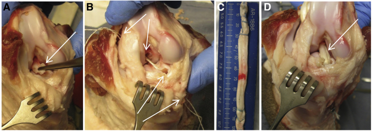
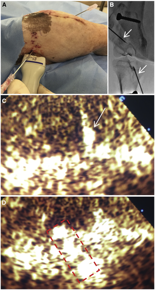
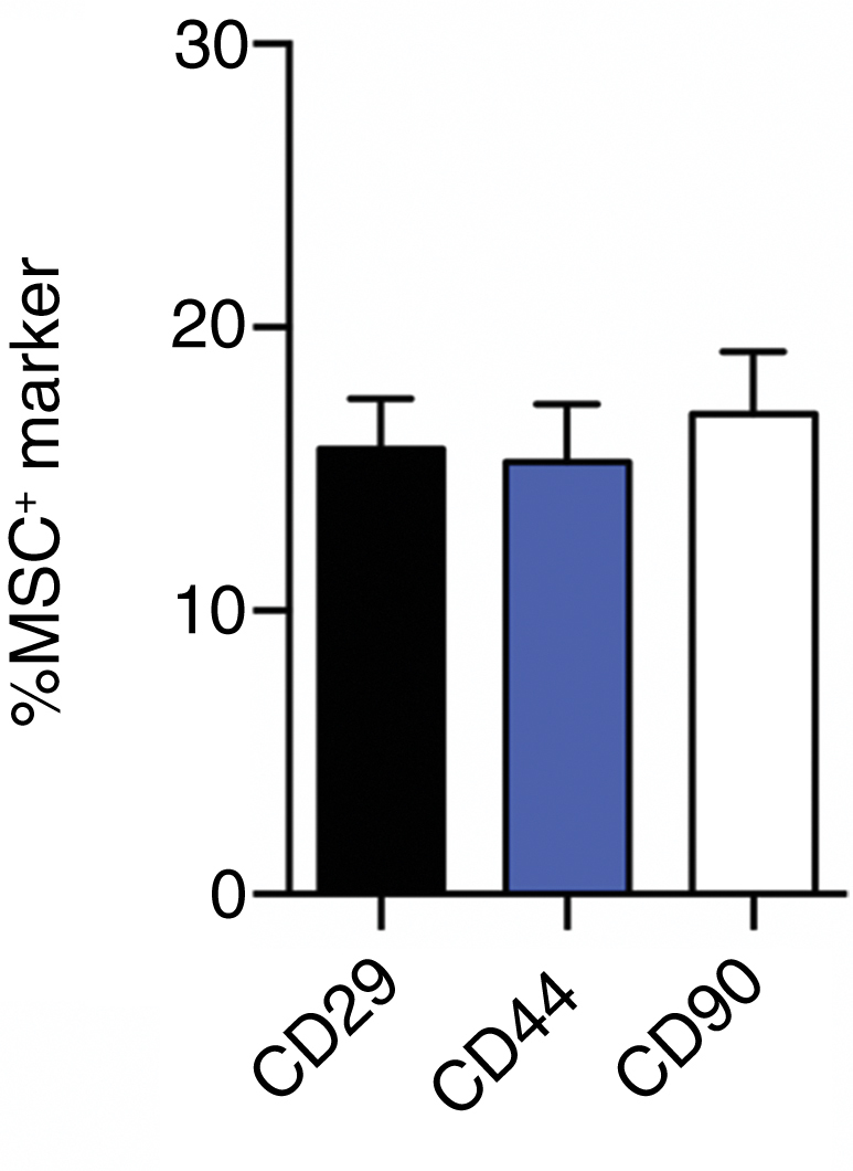
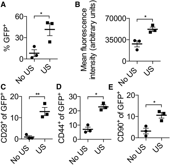
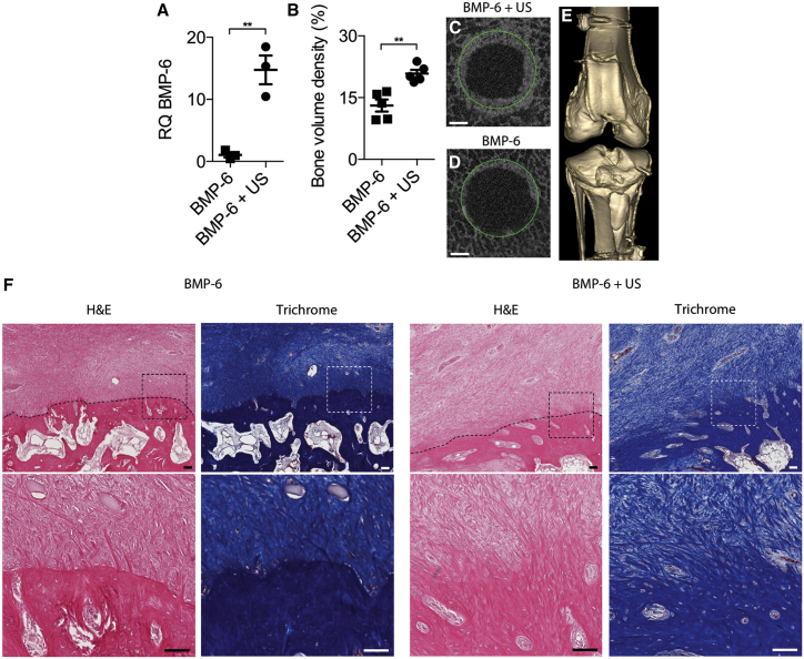
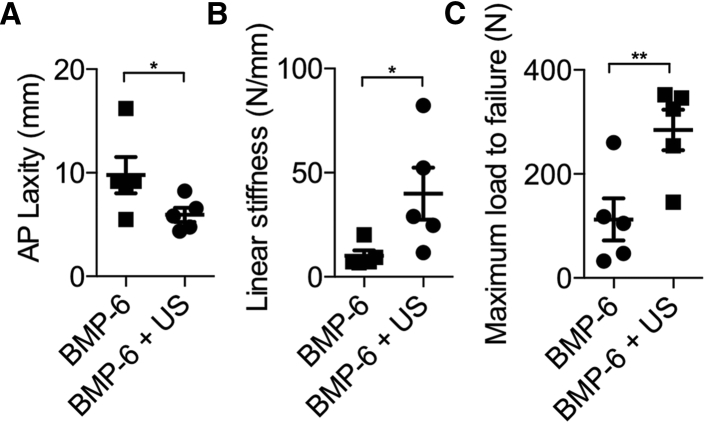
Ligament injuries occur frequently, substantially hindering routine daily activities and sports participation in patients. Surgical reconstruction using autogenous or allogeneic tissues is the gold standard treatment for ligament injuries. Although surgeons routinely perform ligament reconstructions, the integrity of these reconstructions largely depends on adequate biological healing of the interface between the ligament graft and the bone. We hypothesized that localized ultrasound-mediated, microbubble-enhanced therapeutic gene delivery to endogenous stem cells would lead to significantly improved ligament graft integration. To test this hypothesis, an anterior cruciate ligament reconstruction procedure was performed in Yucatan mini-pigs. A collagen scaffold was implanted in the reconstruction sites to facilitate recruitment of endogenous mesenchymal stem cells. Ultrasound-mediated reporter gene delivery successfully transfected 40% of cells recruited to the reconstruction sites. When BMP-6 encoding DNA was delivered, BMP-6 expression in the reconstruction sites was significantly enhanced. Micro-computed tomography and biomechanical analyses showed that ultrasound-mediated BMP-6 gene delivery led to significantly enhanced osteointegration in all animals 8 weeks after surgery. Collectively, these findings demonstrate that ultrasound-mediated gene delivery to endogenous mesenchymal progenitor cells can effectively improve ligament reconstruction in large animals, thereby addressing a major unmet orthopedic need and offering new possibilities for translation to the clinical setting.

Keywords: gene therapy; regenerative medicine; tissue engineering.


Figures

Figure 1
Figure 1
ACL Reconstruction Model in Yucatan Mini-Pig Knee Joints (A) During surgery, the ACL is transected at its tibial and femoral attachments and excised (denoted by arrow). (B) The tibial and femoral tunnels are drilled (arrows), and the required graft length is assessed. (C) The allograft is truncated to the appropriate length and prepared using a running lock suture at each terminus. (D) The allograft (arrow) is inserted into the drilled bone tunnels and tied using sutures to anchor it to cortical screws.
Figure 2
Figure 2
Ultrasound Treatment Setup (A) Injection of a DNA and microbubble mixture into the bone tunnel sites. The ultrasound probe is placed adjacent to the needle for optimal visualization of the injected mixture. (B) Fluoroscopic imaging of needle placement within the bone tunnel sites is shown. Arrows denote the inserted needles. (C) Ultrasound contrast agent imaging during injection of the plasmid and microbubbles mixture is shown. Arrow denotes the inserted needle in the tunnel. (D) Ultrasonographic visualization of the injected mixture (marked with dashed line) within the bone tunnel during sonoporation is shown.
Figure 3
Figure 3
Recruitment of Endogenous Mesenchymal Progenitor Cells to ACL Bone Tunnels Flow cytometry analysis of CD29-, CD44-, and CD90-positive cells from bone tunnel sites 19 days postoperatively (MSC, mesenchymal stem cell). Data are represented as mean ± SEM.
Figure 4
Figure 4
Reporter Gene Expression in Mini-pig Reconstruction Sites following Ultrasound-Mediated Gene Delivery (A) Flow cytometry analysis of the percentage of GFP-positive cells isolated from bone tunnel sites, with or without ultrasound treatment, 5 days after treatment. (B) Mean fluorescence intensity per cell in GFP-positive cells isolated from bone tunnels is shown. (C–E) Percentage of GFP-positive cells positive for CD29 (C), CD44 (D), and CD90 (E). n = 3 per experimental group; *p < 0.05; **p < 0.01; no US, group in which no ultrasound was used; US, group in which ultrasound was used. Data are represented as mean ± SEM.
Figure 5
Figure 5
Ultrasound-Mediated BMP-6 Gene Delivery to Mini-pig ACL Reconstruction Sites (A) BMP-6 gene (relative quantification [RQ]) expression in bone tunnels at ACL reconstruction sites 5 days after treatment. (B) Quantitative analysis of bone formation in reconstruction sites 8 weeks after surgery is shown. (C and D) Representative μCT slices of bone tunnels that received (C) or did not receive (D) ultrasound treatment, obtained 8 weeks after surgery, are shown. Green circles denote the original diameters of the bone tunnels created during surgery. The scale bars represent 1 mm. (E) Fluoroscopic 3D reconstruction of a representative BMP-6 and ultrasound-treated knee joint is shown. (F) Masson’s trichrome and H&E staining of the bone-graft interface 8 weeks after surgery at low magnification (upper) and at high magnification of tissue in the marked squares (lower) is shown. Dotted lines show boundaries between grafts and calcified tissues. The scale bars represent 100 μm. n = 3 per experimental group in the gene expression study; n = 5 per experimental group in the bone formation study; **p < 0.01. Data are represented as mean ± SEM.
Figure 6
Figure 6
Biomechanical Properties of Knee Joints following Ultrasound-Mediated Gene Delivery (A) AP laxity testing of ultrasound-treated and untreated knee joints. (B and C) Linear stiffness (B) and maximum load to failure (C) in implanted grafts at the ACL reconstruction site during tensile failure testing 8 weeks after treatment are shown. AP, anteroposterior; N, Newton; n = 5 per experimental group; *p < 0.05; **p < 0.01. Data are represented as mean ± SEM.

References

    1. Gordon M.D., Steiner M.E. Anterior cruciate ligament injuries. In: Garrick J.G., editor. Orthopaedic Knowledge Update Sports Medicine III. American Academy of Orthopaedic Surgeons; 2004. pp. 169–182.
    1. Yabroudi M.A., Björnsson H., Lynch A.D., Muller B., Samuelsson K., Tarabichi M., Karlsson J., Fu F.H., Harner C.D., Irrgang J.J. Predictors of revision surgery after primary anterior cruciate ligament reconstruction. Orthop. J. Sports Med. 2016;4 2325967116666039. - PMC - PubMed
    1. Smith L., Xia Y., Galatz L.M., Genin G.M., Thomopoulos S. Tissue-engineering strategies for the tendon/ligament-to-bone insertion. Connect. Tissue Res. 2012;53:95–105. - PMC - PubMed
    1. Atesok K., Fu F.H., Wolf M.R., Ochi M., Jazrawi L.M., Doral M.N., Lubowitz J.H., Rodeo S.A. Augmentation of tendon-to-bone healing. J. Bone Joint Surg. Am. 2014;96:513–521. - PubMed
    1. Hofbauer M., Valentin P., Kdolsky R., Ostermann R.C., Graf A., Figl M., Aldrian S. Rotational and translational laxity after computer-navigated single- and double-bundle anterior cruciate ligament reconstruction. Knee Surg. Sports Traumatol. Arthrosc. 2010;18:1201–1207. - PubMed

Publication types

MeSH terms