

The presence of large focal lesions is a strong independent prognostic factor in multiple myeloma
- PMID: 29784643
- PMCID: PMC6034645
- DOI: 10.1182/blood-2018-04-842880
The presence of large focal lesions is a strong independent prognostic factor in multiple myeloma
Abstract
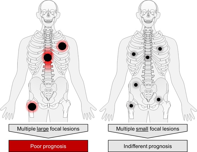
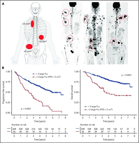
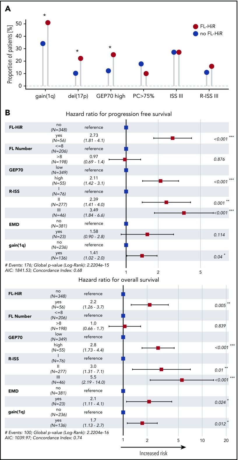
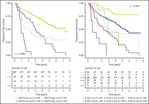
Spatial intratumor heterogeneity is frequently seen in multiple myeloma (MM) and poses a significant challenge for risk classifiers, which rely on tumor samples from the iliac crest. Because biopsy-based assessment of multiple skeletal sites is difficult, alternative strategies for risk stratification are required. Recently, the size of focal lesions (FLs) was shown to be a surrogate marker for spatial heterogeneity, suggesting that data from medical imaging could be used to improve risk stratification approaches. Here, we investigated the prognostic value of FL size in 404 transplant-eligible, newly diagnosed MM patients. Using diffusion-weighted magnetic resonance imaging with background suppression, we identified the presence of multiple large FLs as a strong prognostic factor. Patients with at least 3 large FLs with a product of the perpendicular diameters >5 cm2 were associated with poor progression-free survival (PFS) and overall survival (OS; median, 2.3 and 3.6 years, respectively). This pattern, seen in 13.8% of patients, was independent of the Revised International Staging System (RISS), gene expression profiling (GEP)-based risk score, gain(1q), or extramedullary disease (hazard ratio, 2.7 and 2.2 for PFS and OS in multivariate analysis, respectively). The number of FLs lost its negative impact on outcome after adjusting for FL size. In conclusion, the presence of at least 3 large FL is a feature of high risk, which can be used to refine the diagnosis of this type of disease behavior and as an entry criterion for risk-stratified trials.
© 2018 by The American Society of Hematology.
Conflict of interest statement
Conflict-of-interest disclosure: B.B. is a coinventor on patents and patent applications related to use of gene expression profiling in cancer medicine that have been licensed to Quest Diagnostics. The remaining authors declare no competing financial interests.
Figures
References
-
- Pawlyn C, Morgan GJ. Evolutionary biology of high-risk multiple myeloma. Nat Rev Cancer. 2017;17(9):543-556. - PubMed
-
- Shaughnessy JD Jr, Zhan F, Burington BE, et al. A validated gene expression model of high-risk multiple myeloma is defined by deregulated expression of genes mapping to chromosome 1. Blood. 2007;109(6):2276-2284. - PubMed
-
- Hillengass J, Fechtner K, Weber MA, et al. Prognostic significance of focal lesions in whole-body magnetic resonance imaging in patients with asymptomatic multiple myeloma. J Clin Oncol. 2010;28(9):1606-1610. - PubMed
Publication types
MeSH terms
Grants and funding
LinkOut - more resources
Full Text Sources
Other Literature Sources
Medical
