. 2018 Sep;16(9):1348-1360.
doi: 10.1158/1541-7786.MCR-17-0634. Epub 2018 May 21.

Autophagy, Cell Viability, and Chemoresistance Are Regulated By miR-489 in Breast Cancer

Affiliations

Autophagy, Cell Viability, and Chemoresistance Are Regulated By miR-489 in Breast Cancer

Mithil Soni et al. Mol Cancer Res. 2018 Sep.

Abstract

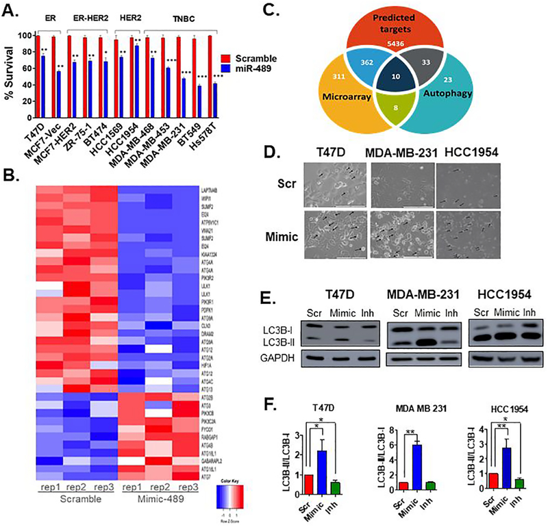
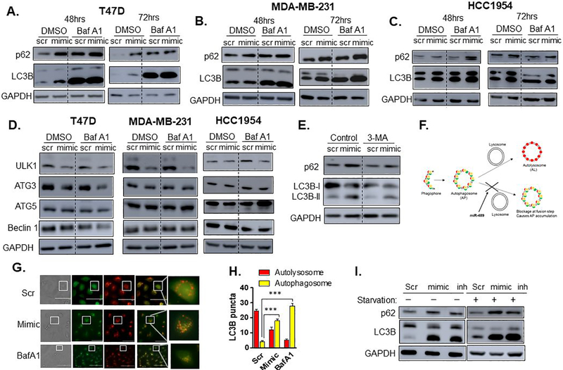
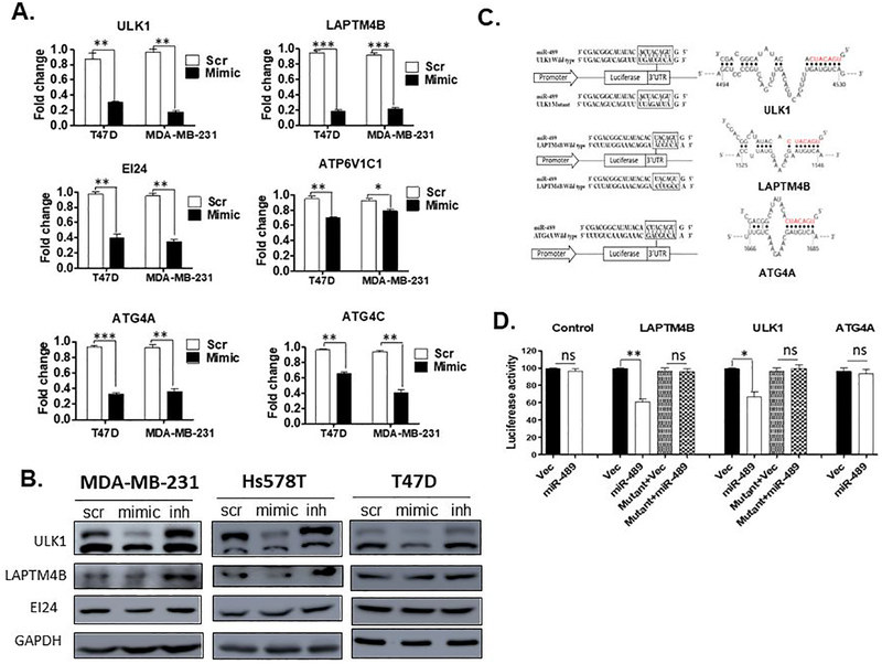
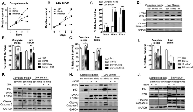
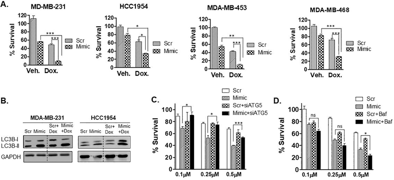
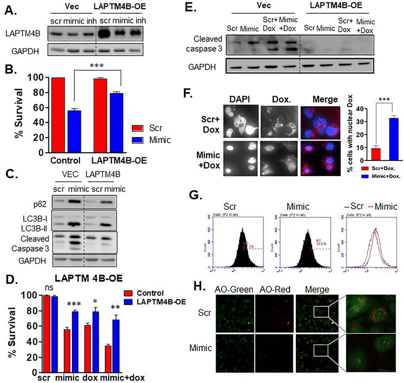
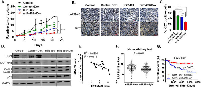
It is postulated that the complexity and heterogeneity in cancer may hinder most efforts that target a single pathway. Thus, discovery of novel therapeutic agents targeting multiple pathways, such as miRNAs, holds promise for future cancer therapy. One such miRNA, miR-489, is downregulated in a majority of breast cancer cells and several drug-resistant breast cancer cell lines, but its role and underlying mechanism for tumor suppression and drug resistance needs further investigation. The current study identifies autophagy as a novel pathway targeted by miR-489 and reports Unc-51 like autophagy activating kinase 1 (ULK1) and lysosomal protein transmembrane 4 beta (LAPTM4B) to be direct targets of miR-489. Furthermore, the data demonstrate autophagy inhibition and LAPTM4B downregulation as a major mechanism responsible for miR-489-mediated doxorubicin sensitization. Finally, miR-489 and LAPTM4B levels were inversely correlated in human tumor clinical specimens, and more importantly, miR-489 expression levels predict overall survival in patients with 8q22 amplification (the region in which LAPTM4B resides).Implications: These findings expand the understanding of miR-489-mediated tumor suppression and chemosensitization in and suggest a strategy for using miR-489 as a therapeutic sensitizer in a defined subgroup of resistant breast cancer patients. Mol Cancer Res; 16(9); 1348-60. ©2018 AACR.


Conflict of interest statement

Disclosure of potential conflicts of interest

The authors have no potential conflict of interest to disclosure.

Figures

Fig. 1.
Fig. 1.. miR-489 inhibits breast cancer cell growth and modulates multiple genes involved in autophagy.
A. MTT based cell proliferation/viability assay measuring viability of indicated breast cancer cell lines at 72hrs post transfection with 28nM scramble control miRNA (Scr) and miR-489 mimic (Mimic). **, p < 0.01; ***, p < 0.001. Data are representative of three independent experiments. B. Heatmap analysis of microarray data for the genes associated with autophagy. Genes were selected based on their differential expression between the scr transfected cells and mimic transfected cells. Red and blue indicate high and low gene expression, respectively. C. Hypergeometric analysis was performed using microarray data and putative targets identified using microRNA target-predicting software. D. Bright-field microscopy images of Scr or Mimic transfected cells. Arrow indicates intracellular endosomal, possibly vacuolar structures. Data are representative of three independent experiments. E. Indicated breast cancer cell lines were transfected with scr, mimic or miR-489 inhibitor (Inh). Protein was collected 72hrs post transfection and subject to western blot analysis of autophagy markers. GAPDH was used as a loading control. F. Quantification of LC3B-I and LC3B-II ratio.
Fig. 2.
Fig. 2.. miR-489 inhibits autophagy mainly by blocking maturation step.
A-C. Bafilomycin A1 blots after reconstituting cells with miR-489. T47D (A.), MDA-MB-231 (B.) and HCC1954 (C.) cell lines were transfected with 28nM scr or mimic, for 48 or 72hrs and p62 and LC3B-II protein expression was assayed by western blot in presence or absence of Bafilomycin A1 (BafA1) (400nM). GAPDH was used as a loading control. D. Western blot analysis of core autophagy genes after 72hrs post transfection. Cells were transfected with 28nM scr or mimic, for 72hrs, and then treated with BafA1 for 4hrs. GAPDH was used as a loading control. E. Western blot analysis of autophagic flux after reconstitution of scr or mimic with or without autophagy inhibitor 3-MA. Cells were transfected with 28nM scr or mimic, for 24hrs, and then treated with 3-MA for 48hrs. Autophagic flux was then analyzed using western blot. GAPDH was used as a loading control. F. Schematic diagram of mCherry-EGFP-LC3B reporter. G. Confocal microscopy of autophagy maturation. MDA-MB-231 cells stably expressing mCherry-EGFP-LC3B fusion protein were transfected with 28nM scr or mimic for 48hrs or Bafilomycin for 4hrs and assayed for co-localization of red and green punta using confocal microscopy. H. Quantitative analysis of red and yellow punta in MDA-MB-231 cells at 48hrs post-transfection of scr or mimic I. miR-489 blocks starvation induced autophagy. MDA-MB-231 cells were transfected with 28nM scr or mimic for 68hrs and treated with EBSS for last 4hrs to induce autophagy. Bafilomycin A1 was used as a control. GAPDH was used as a loading control. **, p < 0.01; ***, p < 0.001. Data are representative of three independent experiments.
Fig. 3.
Fig. 3.. miR-489 directly targets ULK1 and LAMTM4B genes.
A. MDA-MB-231 and T47D cell lines were transfected with 28nM scr or mimic. RNA was isolated 72hrs post transfection and qRT-PCR was performed to examine expression level of indicated genes. Data are means of three replicates ± SEM. B. Western blot showing expression of potential targets upon transfection of 28nM scr, mimic or inh in indicated cell lines. GAPDH was used as a loading control. C. A schematic representation of the target mRNA with putative miR-489 binding site in the 3’ UTR by S fold database, where the seed region is highlighted in red. D. HEK293T cells were co-transfected with miR-489 expressing vector or empty vector and renilla expressing vector for 72hrs. Firefly luciferase was measured for each condition and normalized with renilla luciferase. Normalized luciferase activity was compared with WT-3’UTR and Mutant 3’ UTR of target mRNA. Data are means of three replicates ± SEM.
Fig. 4.
Fig. 4.. miR-489 reduces tumor cell survival and sensitizes tumor cells under starvation by inhibition autophagosome maturation.
MDA-MB-231 cells were transfected with 28nM scr, mimic or inh in complete media (A) or low serum (B). Cell viability assay was performed at indicated time points (0, 24, 28 and 72hrs) using MTT. *, p < 0.05; **, p < 0.01; ***, p < 0.001. Data are means of three replicates ± SEM. C. Relative inhibition by mimic under both conditions at indicated time points. D. MDA-MB-231 cells were transfected with 28nM scr, mimic or inh in complete media or low serum for 72hrs and western blot was performed to investigate expression of cleaved caspase 3 and autophagy markers. GAPDH was used as a loading control. E-J. MDA-MB-231 cells were transfected with 9.3nM scr or mimic in complete media or low serum with or without 3-MA (5mM), siATG5 (50nM) or BafA1 (10nM) treatment and cell viability assay (E, G, I) and western blot analysis (F, H, J) was performed to examine autophagic flux and apoptosis *, p < 0.05; **, p < 0.01; ***, p < 0.001. Data are means of three replicates ± SEM.
Fig. 5.
Fig. 5.. miR-489 sensitizes breast cancer cells to doxorubicin induced cell death by inhibiting doxorubicin induced cytoprotective autophagy.
A. Indicated breast cancer cell lines were transfected with 28nM scr or mimic for 72hrs in presence of doxorubicin (0.75μM) for 48hrs and cell proliferation was measured by MTT assay. Data are representative of three independent experiments. *, p < 0.05; **, p < 0.01; ***, p < 0.001. B. MDA-MB-231 and HCC1954 cells were transfected with scr or mimic for 72hrs in absence or presence of doxorubicin and autophagy marker LC3B-I and LC3B-II were monitored to examine autophagic flux. C. MDA-MB-231 cells were transfected with 9.3nM scr or mimic with or without siATG5 for 24hrs and treated with indicated concentration of doxorubicin for 48hrs and cell proliferation was measured by MTT assay. D. MDA-MB-231 cells were transfected with 9.3nM scr or mimic with for 24hrs and treated with indicated concentration of doxorubicin in presence or absence of Bafilomycin A1 (50nM) for 48hrs and cell proliferation was measured by MTT assay. Data are representative of three independent experiments. *, p < 0.05; **, p < 0.01; ***, p < 0.001.
Fig. 6.
Fig. 6.. miR-489 sensitizes breast cancer cells to doxorubicin partly by targeting LAPTM4B.
A. Western blot indicating stable over expressing LAPTM4B. GAPDH was used as a loading control for western blot analysis. B-C. MDA-MB-231 cells stably expressing LAPTM4B were transfected with 28nM scr or mimic. Cell viability assay (B.) and western blot (C.) was performed 72hrs post transfection to examine autophagic flux and apoptosis. D-E. MDA-MB-231 cells stably expressing LAPTM4B were transfected with 28nM scr or mimic with or without doxorubicin followed by cell viability assay (D.) and western blot analysis (E.) F. Microscopy analysis of subcellular localization of doxorubicin. Confocal microscopy was performed 72hrs after MDA-MB-231 cells were treated with 28nM scr or mimic with doxorubicin. Data are means of three replicates ± SEM. Data are representative of three independent experiments. G. MDA-MB-231 cells were transfected with 28nM scr or mimic and stained with Acridine orange (1mg/ml) for 20min and flow cytometry was performed to examine lysosomal integrity. H. Confocal microscopy of MDA-MB-231 cells after transfection with scr or mimic and staining with Acridine orange (1mug/ml) for 20min. Data are representative of three independent experiments.
Fig. 7.
Fig. 7.. Nanoparticle-delivered miR-489 inhibits tumor growth and sensitized cells against doxorubicin in vivo.
A. miR-489 inhibits tumor growth and sensitized cells against doxorubicin in xenograft animals. After the tumors were palpable, the animals were randomly assigned into four groups (n=5 per group). All animals were injected with miR-489 or control encapsulated in nanoparticle every third day. The treatment starting day was referred to as ‘Day zero’ in the figure. B. IHC analysis revealed reduced expression of LAPTM4B and Ki67 in tumors treated with miR-489 encapsulated nanoparticles. C. Quantification of Ki-67 positive cells in tumors of all four groups. D. Western blot analysis of tumors revealed down regulation of ULK1, LAPTM4B and autophagy inhibition by miR-489. GAPDH was used as a loading control. E. miR-489 and LAPTM4B expression was measured in breast tissues form breast cancer patients (n=14) using qPCR. F. Correlation of miR-489 and its potential target gene expression in primary breast cancers. The linear dependence between miR489 and its potential target genes expression was evaluated by Pearson analysis of a published breast cancer data set (38). G. miR-489 expression predict overall survival of breast cancer patients with 8q22 gain/amplified tumors. Patient survival was estimated using the Kaplan-Meier method and compared with log-rank tests. The Y axis represents the probability of overall survival. *, p < 0.05; **, p < 0.01; ***, p < 0.001.

Similar articles

Cited by

References

    1. Adams BD, Wali VB, Cheng CJ, Inukai S, Booth CJ, Agarwal S, et al. miR-34a Silences c-SRC to Attenuate Tumor Growth in Triple-Negative Breast Cancer. Cancer research 2016;76(4):927–39 doi 10.1158/0008-5472.CAN-15-2321. - DOI - PMC - PubMed
    1. Esparza-Lopez J, Escobar-Arriaga E, Soto-Germes S, Ibarra-Sanchez MJ. Breast Cancer Intra-Tumor Heterogeneity: One Tumor, Different Entities. Revista de investigacion clinica; organo del Hospital de Enfermedades de la Nutricion 2017;69(2):66–76. - PubMed
    1. Iorio MV, Ferracin M, Liu CG, Veronese A, Spizzo R, Sabbioni S, et al. MicroRNA gene expression deregulation in human breast cancer. Cancer research 2005;65(16):7065–70 doi 10.1158/0008-5472.CAN-05-1783. - DOI - PubMed
    1. Patel Y, Shah N, Lee JS, Markoutsa E, Jie C, Liu S, et al. A novel double-negative feedback loop between miR-489 and the HER2-SHP2-MAPK signaling axis regulates breast cancer cell proliferation and tumor growth. Oncotarget 2016;7(14):18295–308 doi 10.18632/oncotarget.7577. - DOI - PMC - PubMed
    1. Lin Y, Liu J, Huang Y, Liu D, Zhang G, Kan H. microRNA-489 Plays an Anti-Metastatic Role in Human Hepatocellular Carcinoma by Targeting Matrix Metalloproteinase-7. Translational oncology 2017;10(2):211–20 doi 10.1016/j.tranon.2017.01.010. - DOI - PMC - PubMed

Publication types

MeSH terms