. 2018 Jun 5;115(23):5950-5955.
doi: 10.1073/pnas.1802501115. Epub 2018 May 21.

VirB8 homolog TraE from plasmid pKM101 forms a hexameric ring structure and interacts with the VirB6 homolog TraD

Affiliations

VirB8 homolog TraE from plasmid pKM101 forms a hexameric ring structure and interacts with the VirB6 homolog TraD

Bastien Casu et al. Proc Natl Acad Sci U S A. .

Abstract

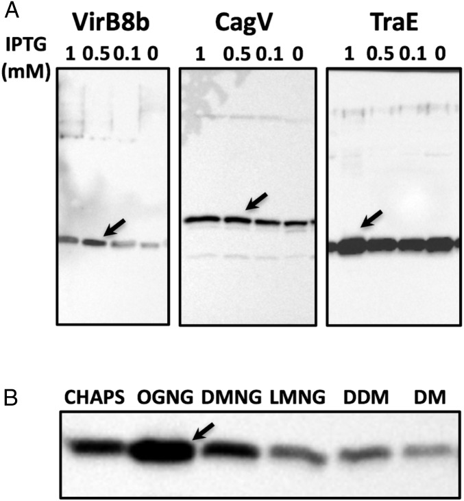
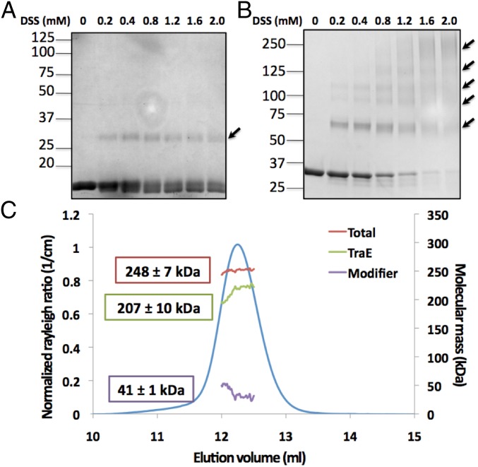
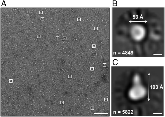
Type IV secretion systems (T4SSs) are multiprotein assemblies that translocate macromolecules across the cell envelope of bacteria. X-ray crystallographic and electron microscopy (EM) analyses have increasingly provided structural information on individual T4SS components and on the entire complex. As of now, relatively little information has been available on the exact localization of the inner membrane-bound T4SS components, notably the mostly periplasmic VirB8 protein and the very hydrophobic VirB6 protein. We show here that the membrane-bound, full-length version of the VirB8 homolog TraE from the plasmid pKM101 secretion system forms a high-molecular-mass complex that is distinct from the previously characterized periplasmic portion of the protein that forms dimers. Full-length TraE was extracted from the membranes with detergents, and analysis by size-exclusion chromatography, cross-linking, and size exclusion chromatography (SEC) multiangle light scattering (MALS) shows that it forms a high-molecular-mass complex. EM and small-angle X-ray scattering (SAXS) analysis demonstrate that full-length TraE forms a hexameric complex with a central pore. We also overproduced and purified the VirB6 homolog TraD and show by cross-linking, SEC, and EM that it binds to TraE. Our results suggest that TraE and TraD interact at the substrate translocation pore of the secretion system.

Keywords: VirB6-like; VirB8-like; conjugation; plasmid; type IV secretion.


Conflict of interest statement

The authors declare no conflict of interest.

Figures

Fig. 1.
Fig. 1.
Overexpression and detergent solubilization of VirB8-like full-length proteins. (A) Western blot analysis with a His-tag–specific antiserum to test the overexpression of VirB8-like proteins using the indicated concentrations of the expression inducer IPTG: Brucella (VirB8b; 30 °C, 6 h), Helicobacter (CagV; 30 °C, 6 h), and pKM101 (TraE; 18 °C, 16 h). (B) Western blot analysis with a His-tag–specific antiserum to test the solubilization of TraE in several detergents (CHAPS, DM, DMNG, LMNG, and OGNG). CHAPS, 3-[(3-cholamidopropyl)dimethylammonio]-1-propanesulfonate; DM, decyl maltoside; DMNG, decyl maltose neopentyl glycol; LMNG, lauryl maltose neopentyl glycol; OGNG, octyl glucose neopentyl glycol. Arrows indicate optimal conditions.
Fig. 2.
Fig. 2.
Analysis of the oligomerization state of TraE. SDS/PAGE analysis of the purified periplasmic domain of TraE at 1 mg/mL (A) and of purified full-length TraE at 1 mg/mL (B) in the absence (0 mM) and presence of increasing concentrations (0.2–2.0 mM) of the cross-linking agent DSS. Proteins in the gels were stained with Coomassie blue dye, and arrows indicate higher molecular-weight complexes formed after cross-linking. (C) Elution profile of the TraE oligomer is shown with the molecular weight estimated by MALS. The molar masses corresponding to the total complex, the TraE oligomer, and the modifier (detergent micelle) throughout the elution peaks are shown.
Fig. 3.
Fig. 3.
EM analysis of the TraE structure. (A) Typical negative-stain micrograph of the TraE complex showing uniform particles of ∼130 Å. (Scale bar, 500 Å.) (B) Representative 2D class averages following alignment, reference-free clustering, and multireference alignment of 1,061 particles. Numbers of particles used for generating each average are shown in the upper left portion of the panels. (Scale bars, 50 Å.) (C) Projections of TraE structures after a low-pass filter at 15 Å and the approximate dimensions are illustrated. (Scale bars, 50 Å.)
Fig. 4.
Fig. 4.
SEC-SAXS analysis of TraE. (A) SEC profile of the TraE sample used for the inline SAXS experiment. mAU, arbitrary units. (B) Normalized pair distribution functions [P(R)] calculated automatically with AutoGNOM. (C) Fit of the theoretical scattering profile for the rigid body model (gray plot) with the experimental SAXS data (black line). A top view (D), side view (E), and down view (F) of the average molecular envelope calculated for TraE (Small Angle Scattering Biological Databank SASDB75) are shown. The approximate envelope dimensions are illustrated.
Fig. 5.
Fig. 5.
SEC and cross-linking analysis of TraD and the TraD–TraE complex. (A) SEC profile of the TraD–TraE complex showing an apparent molecular mass of 200 kDa. SDS/PAGE and Western blot analysis of the SEC peak fraction with TraE-specific antiserum and His-tag–specific antiserum to detect N-terminally His-tagged TraD (His6-TraD), SDS/PAGE, and Coomassie blue staining shows the purity of the complex. mAU, arbitrary units. (B) Purified His6-TraD was incubated with varying concentrations of DSS, and cross-linking products were detected after SDS/PAGE and Western blotting with His-tag–specific antiserum. (C) Purified His6-TraD–TraE complex was incubated with varying concentrations of DSS, and cross-linking products were detected after SDS/PAGE and Western blotting with antiserum specific for His6-TraD. (D) Purified His6-Tra–TraE complex was incubated with varying concentrations of DSS, and cross-linking products were detected after SDS/PAGE and Western blotting with TraE-specific antiserum. Arrows indicate higher molecular-weight complexes formed after cross-linking.
Fig. 6.
Fig. 6.
EM analysis of the TraD–TraE complex structure. (A) Typical negative-stain micrograph of the TraD–TraE complex showing uniform particles with dimensions of ∼60 Å and ∼100 Å, respectively. (Scale bar, 500 Å.) (B and C) Representative 2D class averages following alignment, reference-free clustering, and multireference alignment of 84,236 particles. Numbers of particles used for generating each average are shown in the bottom left portion of the panels, and the approximate dimensions are illustrated. (Scale bars, 30 Å.)

Similar articles

Cited by

References

    1. Trokter M, Felisberto-Rodrigues C, Christie PJ, Waksman G. Recent advances in the structural and molecular biology of type IV secretion systems. Curr Opin Struct Biol. 2014;27:16–23. - PMC - PubMed
    1. Chandran Darbari V, Waksman G. Structural biology of bacterial type IV secretion systems. Annu Rev Biochem. 2015;84:603–629. - PubMed
    1. Burkinshaw BJ, Strynadka NC. Assembly and structure of the T3SS. Biochim Biophys Acta. 2014;1843:1649–1663. - PubMed
    1. Winans SC, Walker GC. Conjugal transfer system of the IncN plasmid pKM101. J Bacteriol. 1985;161:402–410. - PMC - PubMed
    1. Fronzes R, Christie PJ, Waksman G. The structural biology of type IV secretion systems. Nat Rev Microbiol. 2009;7:703–714. - PMC - PubMed

Publication types

MeSH terms