. 2018 Jun 5;115(23):E5298-E5306.
doi: 10.1073/pnas.1722633115. Epub 2018 May 21.

Xenoprotein engineering via synthetic libraries

Affiliations

Xenoprotein engineering via synthetic libraries

Zachary P Gates et al. Proc Natl Acad Sci U S A. .

Abstract

Chemical methods have enabled the total synthesis of protein molecules of ever-increasing size and complexity. However, methods to engineer synthetic proteins comprising noncanonical amino acids have not kept pace, even though this capability would be a distinct advantage of the total synthesis approach to protein science. In this work, we report a platform for protein engineering based on the screening of synthetic one-bead one-compound protein libraries. Screening throughput approaching that of cell surface display was achieved by a combination of magnetic bead enrichment, flow cytometry analysis of on-bead screens, and high-throughput MS/MS-based sequencing of identified active compounds. Direct screening of a synthetic protein library by these methods resulted in the de novo discovery of mirror-image miniprotein-based binders to a ∼150-kDa protein target, a task that would be difficult or impossible by other means.

Keywords: D-protein; flow cytometry; mirror-image miniprotein; protein engineering; xenoprotein.


Conflict of interest statement

The authors declare no conflict of interest.

Figures

Fig. 1.
Fig. 1.
An approach to identifying binders from synthetic protein libraries. Thirty-micrometer beads displaying xenoprotein variants are prepared by a combination of stepwise SPPS and on-bead folding. The resulting beads are incubated with a protein target bearing a fluorescent label (red star), and beads displaying functional xenoprotein variants are isolated by fluorescence-activated sorting (FACS). The sequences of xenoproteins contained on sorted beads are then determined by de novo MS/MS-based peptide sequencing.
Fig. 2.
Fig. 2.
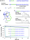
EETI-II is a robust scaffold for the display of chemical diversity. (A) Amino acid sequences of EETI-II and a synthetic library scaffold based on mirror-image EETI-II. d-amino acids are in lowercase; cysteine residues (all in disulfide form) are underlined. Residues corresponding to the trypsin-binding loop are shown in green. (B) Cartoon rendering of the EETI-II molecule, based on Protein Data Bank ID code 1W7Z. The trypsin-binding loop is shown in green, and the three disulfide bonds as yellow sticks. (C) LC-MS data showing the spontaneous oxidative folding of a synthetic EETI-II variant with a nonnative trypsin-binding loop sequence. A loss of 6 Da was observed upon treatment with soluble redox buffer (SI Appendix, section 3), consistent with the formation of three disulfide bonds. Calculated and observed monoisotopic masses are indicated. (D) Stability of l- and d-forms of two engineered EETI-II variants to proteinase K. The fraction of intact protein remaining at each time point was determined by LC-MS–based quantitation (average of two measurements).
Fig. 3.
Fig. 3.
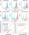
Flow cytometry is amenable to monitoring on-bead binding assays. (A) Fluorescence histograms showing the contrast between library beads (cyan) and StrepTag II beads (maroon) achieved by the use of different fluorescent SA reagents. (B) Fluorescence histograms showing protein target-dependent binding to beads functionalized with the indicated peptide ligands. (C) A plot of the fluorescence means obtained by treatment of biotin-functionalized beads (four different bead loadings) with increasing concentrations of SA-APC. Gly-functionalized beads served as a negative control. (D) A plot of the fluorescence means obtained by treatment of IgG1 binder-functionalized beads with increasing concentrations of a fixed molar ratio of SA-APC and biotinylated polyclonal IgG1. Library beads served as a negative control.
Fig. 4.
Fig. 4.
Folded EETI-II can be prepared on beads. (A) Fluorescence histograms showing the contrast between library beads (cyan) and either EETI-II beads (maroon, Right) or trypsin-binding loop beads (maroon, Left) after incubation with a mixture of SA-APC (50 nM) and biotinylated trypsin (100 nM). (B) Fluorescence means obtained by treatment of the same samples with a fixed concentration of SA-APC (50 nM) and one of two different concentrations of biotinylated trypsin. Trypsin-dependent binding was observed for the EETI-II beads only. (C) LC-MS data showing the spontaneous oxidative folding of EETI-II while bound to beads. (D) LC-MS data showing the spontaneous oxidative folding of an analog of the engineered EETI-II variant 2.5F, while bound to beads; † denotes a three-disulfide-containing product and * denotes a two-disulfide-containing product. For C and D, the indicated mass values correspond to monoisotopic masses.
Fig. 5.
Fig. 5.
(AC) Strategy for the preparation of a mirror-image EETI-II-based library.
Fig. 6.
Fig. 6.
Replicate preparations of identified active library beads exhibit reproducible on-bead binding. Comparable 12CA5-dependent binding was observed for beads functionalized with either (A) HA epitope or (B) putative 12CA5-binding MIMs.
Fig. 7.
Fig. 7.
Orthogonal binding assays confirm the activity of 12CA5-binding MIMs. (A) LC-MS analysis of representative biotinylated 12CA5-binding MIM 1. (B) BLI assay, showing association of exogenous 12CA5 to immobilized MIM 1. (C) FP competition binding assay, showing the displacement of fluorescent HA epitope by unlabeled HA epitope (red) and MIM 1 (black).

References

    1. Wang L, Schultz PG. Expanding the genetic code. Angew Chem Int Ed Engl. 2004;44:34–66. - PubMed
    1. Jackson JC, Duffy SP, Hess KR, Mehl RA. Improving nature’s enzyme active site with genetically encoded unnatural amino acids. J Am Chem Soc. 2006;128:11124–11127. - PubMed
    1. Ugwumba IN, et al. Improving a natural enzyme activity through incorporation of unnatural amino acids. J Am Chem Soc. 2011;133:326–333. - PubMed
    1. Liu CC, et al. Protein evolution with an expanded genetic code. Proc Natl Acad Sci USA. 2008;105:17688–17693. - PMC - PubMed
    1. Liu CC, et al. Evolution of proteins with genetically encoded “chemical warheads”. J Am Chem Soc. 2009;131:9616–9617. - PMC - PubMed

Publication types