

SIRT1 inhibits rheumatoid arthritis fibroblast-like synoviocyte aggressiveness and inflammatory response via suppressing NF-κB pathway
- PMID: 29784872
- PMCID: PMC6013706
- DOI: 10.1042/BSR20180541
SIRT1 inhibits rheumatoid arthritis fibroblast-like synoviocyte aggressiveness and inflammatory response via suppressing NF-κB pathway
Expression of concern in
-
Expression of Concern: SIRT1 inhibits rheumatoid arthritis fibroblast-like synoviocyte aggressiveness and inflammatory response via suppressing NF-κB pathway.Biosci Rep. 2023 Aug 31;43(8):BSR-2018-0541_EOC. doi: 10.1042/BSR-2018-0541_EOC. Biosci Rep. 2023. PMID: 37650880 Free PMC article. No abstract available.
Abstract
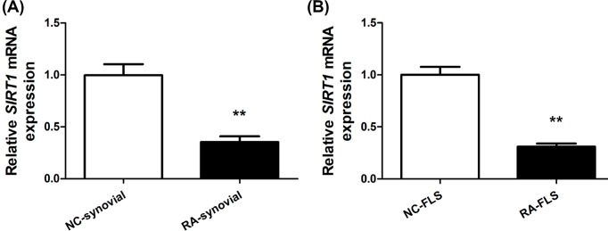
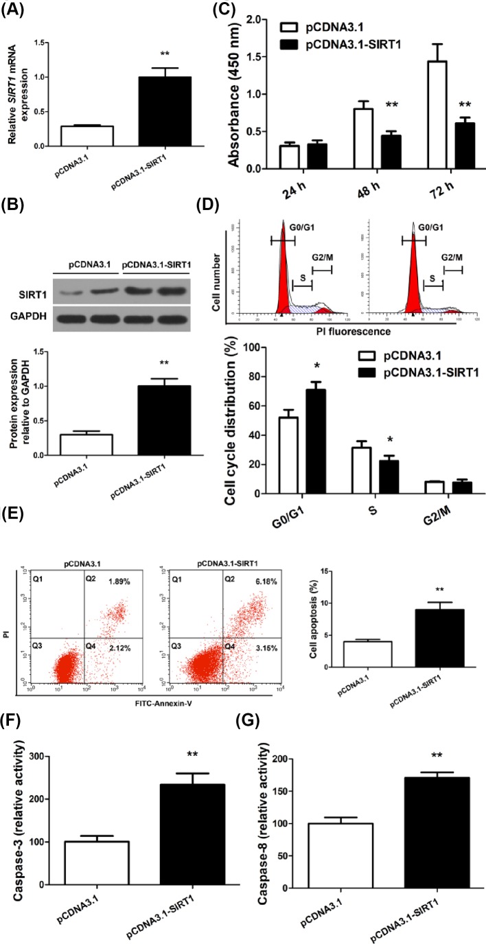
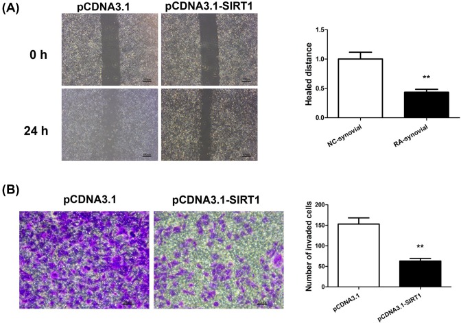
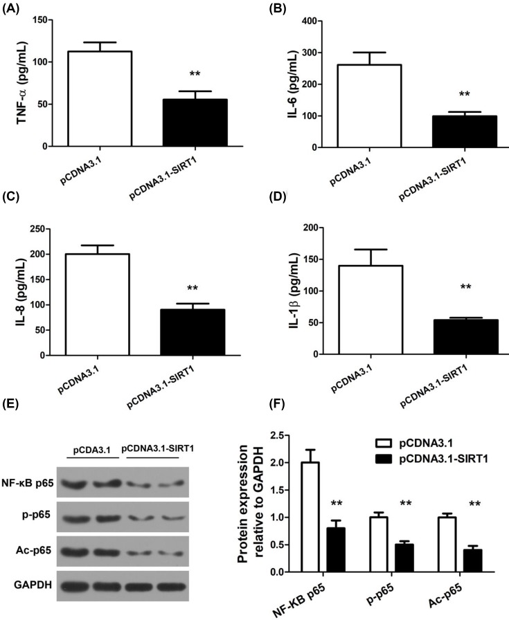
Rheumatoid arthritis (RA) is an autoimmune disease of the joints characterized by synovial hyperplasia and chronic inflammation. Fibroblast-like synoviocytes (FLS) play a central role in RA initiation, progression, and perpetuation. Prior studies showed that sirtuin 1 (SIRT1), a deacetylase participating in a broad range of transcriptional and metabolic regulations, may impact cell proliferation and inflammatory responses. However, the role of SIRT1 in RA-FLS was unclear. Here, we explored the effects of SIRT1 on the aggressiveness and inflammatory responses of cultured RA-FLS. SIRT1 expression was significantly lower in synovial tissues and FLS from RA patients than from healthy controls. Overexpression of SIRT1 significantly inhibited RA-FLS proliferation, migration, and invasion. SIRT1 overexpression also significantly increased RA-FLS apoptosis and caspase-3 and -8 activity. Focusing on inflammatory phenotypes, we found SIRT1 significantly reduced RA-FLS secretion of TNF-α, IL-6, IL-8, and IL-1β. Mechanistic studies further revealed SIRT1 suppressed NF-κB pathway by reducing p65 protein expression, phosphorylation, and acetylation in RA-FLS. Our results suggest SIRT1 is a key regulator in RA pathogenesis by suppressing aggressive phenotypes and inflammatory response of FLS. Enhancing SIRT1 expression or function in FLS could be therapeutic beneficial for RA by inhibiting synovial hyperplasia and inflammation.
Keywords: NF-κB; fibroblast-like synoviocytes; inflammation; rheumatoid arthritis; sirtuin 1.
© 2018 The Author(s).
Conflict of interest statement
All the authors mutually agree to submit our manuscript titled “SIRT1 inhibits rheumatoid arthritis fibroblast-like synoviocyte aggressiveness and inflammatory response via suppressing NF-κB pathway” to
Figures
Similar articles
-
[Mechanism of 4-methylcatechol in inhibiting fibroblast-like synoviocyte migration and suppressing inflammatory responses in treatment of rheumatoid arthritis].Zhongguo Xiu Fu Chong Jian Wai Ke Za Zhi. 2025 Aug 15;39(8):1051-1060. doi: 10.7507/1002-1892.202503013. Zhongguo Xiu Fu Chong Jian Wai Ke Za Zhi. 2025. PMID: 40830133 Free PMC article. Chinese.
-
IRF9 Affects the TNF-Induced Phenotype of Rheumatoid-Arthritis Fibroblast-Like Synoviocytes via Regulation of the SIRT-1/NF-κB Signaling Pathway.Cells Tissues Organs. 2020;209(2-3):110-119. doi: 10.1159/000508405. Epub 2020 Aug 7. Cells Tissues Organs. 2020. PMID: 32772027
-
Triptolide decreases rheumatoid arthritis fibroblast-like synoviocyte proliferation, invasion, inflammation and presents a therapeutic effect in collagen-induced arthritis rats via inactivating lncRNA RP11-83J16.1 mediated URI1 and β-catenin signaling.Int Immunopharmacol. 2021 Oct;99:108010. doi: 10.1016/j.intimp.2021.108010. Epub 2021 Aug 3. Int Immunopharmacol. 2021. PMID: 34358861
-
E3 ubiquitin ligase gene BIRC3 modulates TNF-induced cell death pathways and promotes aberrant proliferation in rheumatoid arthritis fibroblast-like synoviocytes.Front Immunol. 2024 Sep 5;15:1433898. doi: 10.3389/fimmu.2024.1433898. eCollection 2024. Front Immunol. 2024. PMID: 39301019 Free PMC article. Review.
-
Unveiling the Therapeutic Potential: Targeting Fibroblast-like Synoviocytes in Rheumatoid Arthritis.Expert Rev Mol Med. 2025 Jun 5;27:e18. doi: 10.1017/erm.2025.11. Expert Rev Mol Med. 2025. PMID: 40468839 Free PMC article. Review.
Cited by
-
Antioxidant Activity and the Potential Mechanism of the Fruit From Ailanthus altissima Swingle.Front Vet Sci. 2021 Dec 13;8:784898. doi: 10.3389/fvets.2021.784898. eCollection 2021. Front Vet Sci. 2021. PMID: 34966812 Free PMC article.
-
The synovial fluid fibroblast-like synoviocyte: A long-neglected piece in the puzzle of rheumatoid arthritis pathogenesis.Front Immunol. 2022 Aug 5;13:942417. doi: 10.3389/fimmu.2022.942417. eCollection 2022. Front Immunol. 2022. PMID: 35990693 Free PMC article. Review.
-
Sirtuin family in autoimmune diseases.Front Immunol. 2023 Jul 6;14:1186231. doi: 10.3389/fimmu.2023.1186231. eCollection 2023. Front Immunol. 2023. PMID: 37483618 Free PMC article. Review.
-
Decreased SIRT1 expression in the peripheral blood of patients with Graves' disease.J Endocrinol. 2020 Aug;246(2):161-173. doi: 10.1530/JOE-19-0501. J Endocrinol. 2020. PMID: 32485674 Free PMC article.
-
miR-223: An Effective Regulator of Immune Cell Differentiation and Inflammation.Int J Biol Sci. 2021 Jun 4;17(9):2308-2322. doi: 10.7150/ijbs.59876. eCollection 2021. Int J Biol Sci. 2021. PMID: 34239357 Free PMC article. Review.
References
-
- Brooks P.M. (2001) Report of the sixth joint WHO/ILAR task force meeting on rheumatic diseases, 16 January 2000, Geneva, Switzerland. J. Rheumatol. 28, 2540–2543 - PubMed
-
- Yu C., Jin S.Y. and Li M.T. (2018) Current status of rheumatoid arthritis registries and prevalence of remission. Zhonghua Nei Ke Za Zhi 57, 175–178 - PubMed
Publication types
MeSH terms
Substances
LinkOut - more resources
Full Text Sources
Other Literature Sources
Medical
Research Materials
Miscellaneous