. 2018 Jul 6;293(27):10707-10717.
doi: 10.1074/jbc.RA117.001698. Epub 2018 May 21.

A human-specific, truncated α7 nicotinic receptor subunit assembles with full-length α7 and forms functional receptors with different stoichiometries

Affiliations

A human-specific, truncated α7 nicotinic receptor subunit assembles with full-length α7 and forms functional receptors with different stoichiometries

Matías Lasala et al. J Biol Chem. .

Abstract

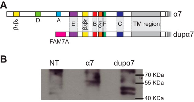
The cholinergic α7 nicotinic receptor gene, CHRNA7, encodes a subunit that forms the homopentameric α7 receptor, involved in learning and memory. In humans, exons 5-10 in CHRNA7 are duplicated and fused to the FAM7A genetic element, giving rise to the hybrid gene CHRFAM7A Its product, dupα7, is a truncated subunit lacking part of the N-terminal extracellular ligand-binding domain and is associated with neurological disorders, including schizophrenia, and immunomodulation. We combined dupα7 expression on mammalian cells with patch clamp recordings to understand its functional role. Transfected cells expressed dupα7 protein, but they exhibited neither surface binding of the α7 antagonist α-bungarotoxin nor responses to acetylcholine (ACh) or to an allosteric agonist that binds to the conserved transmembrane region. To determine whether dupα7 assembles with α7, we generated receptors comprising α7 and dupα7 subunits, one of which was tagged with conductance substitutions that report subunit stoichiometry and monitored ACh-elicited channel openings in the presence of a positive allosteric α7 modulator. We found that α7 and dupα7 subunits co-assemble into functional heteromeric receptors, which require at least two α7 subunits for channel opening, and that dupα7's presence in the pentameric arrangement does not affect the duration of the potentiated events compared with that of α7. Using an α7 subunit mutant, we found that activation of (α7)2(dupα7)3 receptors occurs through ACh binding at the α7/α7 interfacial binding site. Our study contributes to the understanding of the modulation of α7 function by the human specific, duplicated subunit, associated with human disorders.

Keywords: Cys-loop receptor; channel activation; ion channel; nicotinic acetylcholine receptors (nAChR); patch clamp; receptor structure-function; single-channel recording; α7 receptor.


Conflict of interest statement

The authors declare that they have no conflicts of interest with the contents of this article

Figures

Figure 1.
Figure 1.
dupα7 subunit. A, diagram of α7 and dupα7 subunits, showing the loops that form the ACh-binding site (loops A–F); loops at the coupling region (β1β2, Cys, and β8β9 loops), the transmembrane (TM) region, and the FAM7A part of dupα7 at the N-terminal domain. B, expression of dupα7 on BOSC-23 cells. Lysates of BOSC-23 cells transfected with α7 or dupα7 cDNA were probed by Western blotting for the subunit protein. NT, corresponds to lysates from nontransfected cells.
Figure 2.
Figure 2.
α-BTX labeling of BOSC cells transfected with α7 and/or dupα7. Cells were transfected with α7 cDNA, dupα7 cDNA, or with the 1:3 α7/dupα7 cDNA combination. Mock transfected cells correspond to cells transfected with irrelevant plasmid DNA. The total amount of DNA per transfection was normalized with the addition of irrelevant plasmid DNA. A, representative confocal microscopy images showing the membrane fluorescence signal generated in transfected cells stained with Alexa Fluor 488-labeled α-BTX. A zoomed image is included at the lower right corner of each panel. Scale bars correspond to 40 μm for the nonzoomed images and 20 μm for the zoomed images. B, scatter plot of fluorescence intensity in the region of interest (ROI) of individual transfected cells. Results are expressed as mean ± S.D. (n = 15). Different letters (a–c) denote statistically significant differences among groups (p < 0.0001; Sidak's multiple comparisons test).
Figure 3.
Figure 3.
Functional responses of α7 or dupα7. BOSC-23 cells were transfected with α7 or dupα7 cDNAs together with NACHO and Ric-3 as described under “Experimental procedures.” A, representative single-channel currents activated by 100 μm ACh from cells transfected with α7. Typical open and burst duration histograms are shown. B, representative traces of ACh-elicited single-channel currents in the presence of 1 μm PNU-120596 from cells transfected with α7 (top) or dupα7 cDNAs (bottom). Typical open and cluster duration histograms are shown for α7. C, single channel currents activated by the allosteric agonist, 4BP-TQS (50 μm) from cells transfected with α7 (top) or dupα7 cDNAs (bottom). Typical open and cluster duration histograms are shown for α7. Membrane potential: −70 mV. Filter: 9 kHz for ACh and 3 kHz for ACh and PNU-120596 or 4BP-TQS. Openings are shown as upward deflections. In all histograms, dashed gray lines correspond to individual components, and black lines correspond to the sum of components. D, macroscopic currents activated by 100 μm ACh or 30 μm 4BP-TQS from cells transfected with α7 or dupα7 cDNAs. No currents from dupα7-expressing cells were elicited by the allosteric agonist.
Figure 4.
Figure 4.
Electrical fingerprinting for α7/α7LC and dupα7/α7LC heteromeric receptors. Top, models showing the different amino acid residues at the intracellular region for α7 and α7LC. Left panels, representative single-channel currents activated by 100 μm ACh + 1 μm PNU-120596 from cells expressing α7 (A), α7LC (B), α7 + α7LC (C), dupα7 (D), dupα7 + α7LC (E), and dupα7LC + α7 (F). The traces for the mixed subunit conditions are excerpts from the same recording. Membrane potential: −70 mV. Filter: 3 kHz. Channel openings are shown as upward deflections. The dashed lines indicate the amplitude of the different amplitude classes. Right panels: typical amplitude histograms for a whole recording constructed with events longer than 1 ms are shown with the fitted components.
Figure 5.
Figure 5.
Amplitude classes for the α7/α7LC and dupα7/α7LC combinations. A and B, analysis of the amplitude classes for cells transfected with α7LC and α7 (A) or dupα7 (B) cDNAs. Left: A, plot of mean current amplitude against the number of α7LC subunits in the pentameric arrangement for the α7/α7LC combination. The fitted slope by least-squares method is 1.81 ± 0.06 pA/LC subunit. Data are plotted as mean ± S.D. of n = 7 for amplitude class of 10, n = 10 for amplitude classes of 8 and 6 pA, and n = 8 for the amplitude class of 4 pA. B, plot of mean current amplitude against the number of α7LC subunits in the pentameric arrangement for the α7LC/dupα7 combination. The fitted slope by the least-squares method is 1.63 ± 0.3 pA/LC subunit. Data are plotted as mean ± S.D. of n = 15 for amplitude class of 6 and n = 3 for the amplitude class of 4 pA. Right, representative dot plots showing the distribution of clusters as a function of their mean amplitude. Recordings were obtained from cells transfected with α7/α7LC (A) or dupα7/α7LC (B). Each plot corresponds to a single recording, and each point, to a single cluster. The number of amplitude classes was determined by the X-means algorithm included in the QuB software.
Figure 6.
Figure 6.
Requirements for (α7)2(dupα7)3 channel activation. A, diagram showing the α7-binding site for ACh. Mutations were introduced at the complementary face (W55T). B, possible subunit arrangements of receptors containing three dupα7 and two α7 subunits. The arrow shows the functional α7/α7 interfacial-binding site. C, representative single-channel recordings in the presence of 100 μm ACh + 1 μm PNU-120596 showing the lack of channel activity in cells transfected with dupα7 and α7W55T cDNA. The black arrows show possible ACh-binding sites in which the complementary face is provided by dupα7. Membrane potential: −70 mV. Filter: 3 kHz.
Figure 7.
Figure 7.
Superimposed molecular models of dupα7 and α7. A, structural alignment of extracellular domains of two adjacent α7/α7 and dupα7/dupα7 subunits. Human α7 structural model was created by homology modeling based on the structure of the α7-AChBP chimera (PDB code 5AFM) and the 3D model of dupα7 was generated by the I-TASSER server (see “Experimental procedures”). The α7 subunits are shown in gray. In dupα7 subunits the region corresponding to FAM7A is shown in red and that corresponding to α7 in blue. Binding and interface loops present in both molecules are indicated with blue letters and those present in α7 but absent in dupα7 with black letters. B, alignment of the α7 and dupα7 sequences (accessions numbers CAD88995 and NP_647536). The α7 sequence does not include the signal peptide. The sequences are identical after amino acid residue 95 of α7. dupα7 sequence corresponding to the FAM7A region is in red. Residues for the six binding loops (A–F) and loops at the coupling region are indicated with black and gray lines, respectively. Aromatic residues reported as essential for α7 agonist response are in gray boxes.

References

    1. Corringer P.-J., Poitevin F., Prevost M. S., Sauguet L., Delarue M., and Changeux J.-P. (2012) Structure and pharmacology of pentameric receptor channels: from bacteria to brain. Structure 20, 941–956 10.1016/j.str.2012.05.003 - DOI - PubMed
    1. Sauguet L., Shahsavar A., Poitevin F., Huon C., Menny A., Nemecz À., Haouz A., Changeux J.-P., Corringer P.-J., and Delarue M. (2014) Crystal structures of a pentameric ligand-gated ion channel provide a mechanism for activation. Proc. Natl. Acad. Sci. U.S.A. 111, 966–971 10.1073/pnas.1314997111 - DOI - PMC - PubMed
    1. Morales-Perez C. L., Noviello C. M., and Hibbs R. E. (2016) X-ray structure of the human α4β2 nicotinic receptor. Nature 538, 411–415 10.1038/nature19785 - DOI - PMC - PubMed
    1. Lendvai B., Kassai F., Szájli A., and Némethy Z. (2013) α7 Nicotinic acetylcholine receptors and their role in cognition. Brain Res. Bull. 93, 86–96 10.1016/j.brainresbull.2012.11.003 - DOI - PubMed
    1. Corradi J., and Bouzat C. (2016) Understanding the bases of function and modulation of α7 nicotinic receptors: implications for drug discovery. Mol. Pharmacol. 90, 288–299 10.1124/mol.116.104240 - DOI - PubMed

Publication types

MeSH terms

Associated data